. 2021 Dec;15(4):581-594.
doi: 10.1007/s12079-021-00627-1. Epub 2021 Jun 1.

Tsukushi is essential for the formation of the posterior semicircular canal that detects gait performance

Affiliations

Tsukushi is essential for the formation of the posterior semicircular canal that detects gait performance

Toru Miwa et al. J Cell Commun Signal. 2021 Dec.

Abstract

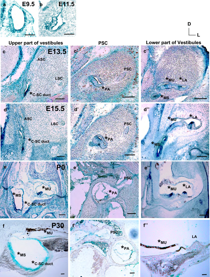
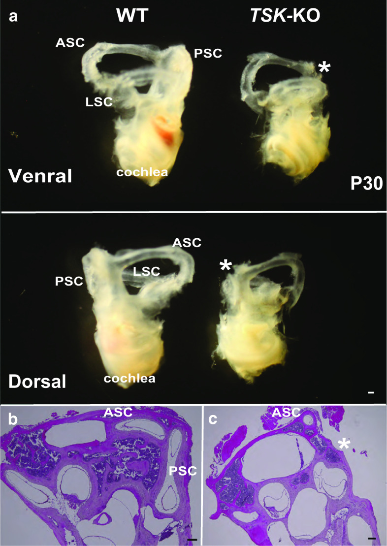
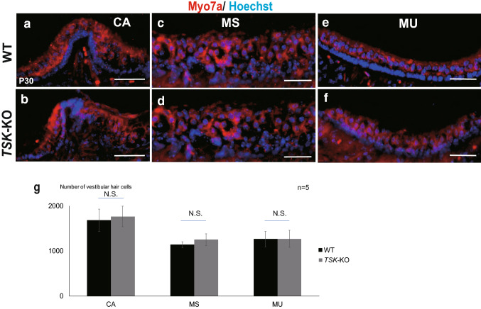
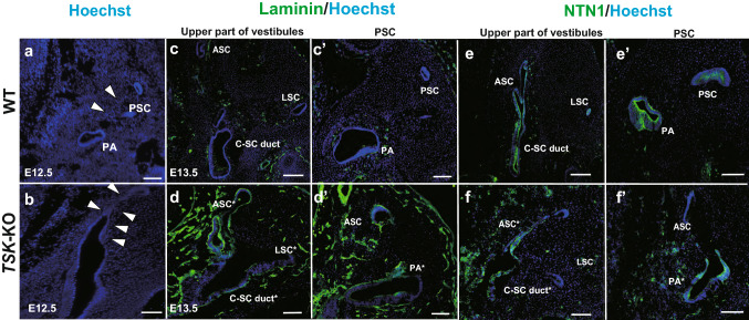
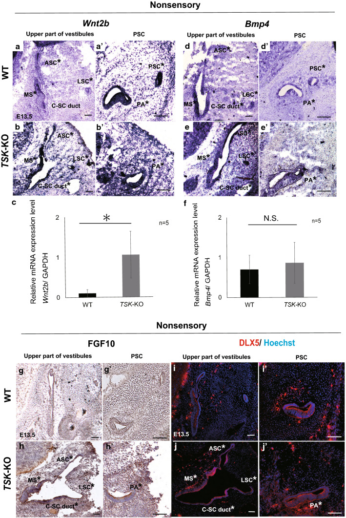
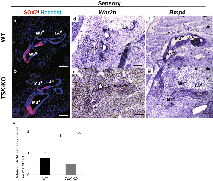
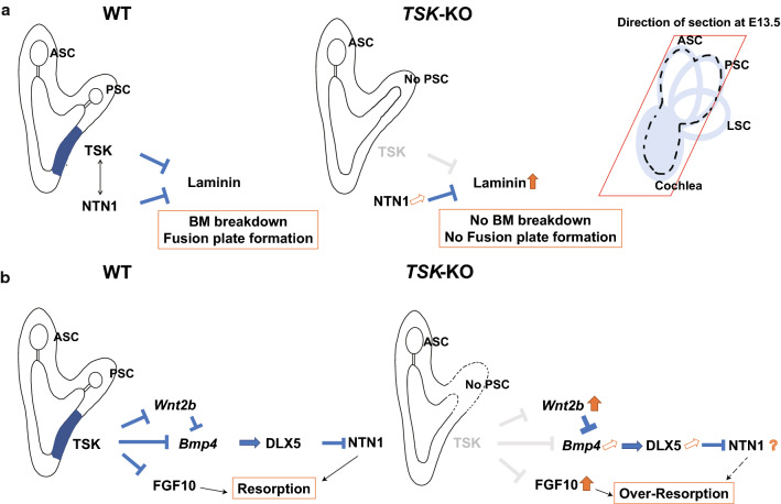
Tsukushi is a small, leucine-rich repeat proteoglycan that interacts with and regulates essential cellular signaling cascades in the chick retina and murine subventricular zone, hippocampus, dermal hair follicles, and the cochlea. However, its function in the vestibules of the inner ear remains unknown. Here, we investigated the function of Tsukushi in the vestibules and found that Tsukushi deficiency in mice resulted in defects in posterior semicircular canal formation in the vestibules, but did not lead to vestibular hair cell loss. Furthermore, Tsukushi accumulated in the non-prosensory and prosensory regions during the embryonic and postnatal developmental stages. The downregulation of Tsukushi altered the expression of key genes driving vestibule differentiation in the non-prosensory regions. Our results indicate that Tsukushi interacts with Wnt2b, bone morphogenetic protein 4, fibroblast growth factor 10, and netrin 1, thereby controlling semicircular canal formation. Therefore, Tsukushi may be an essential component of the molecular pathways regulating vestibular development.

Keywords: Bmp4; FGF10; Posterior semicircular canal; Tsukushi; Vestibule; Wnt2b.


Conflict of interest statement

The authors declare no conflicts of interest.

Figures

Fig. 1
Fig. 1
Spatiotemporal expression of TSK in the inner ear of TSK heterozygous mice. a, b At E9.5 and E11.5, TSK-LacZ-expressing cells were localized in the whole epithelium of the otocyst. cf Upper part of the vestibules at E13.5 (c), E15.5 (d), P0 (e), and P30 (f). TSK-LacZ-expressing cells were localized at the C-SC duct (indicated with asterisks). At P0 and P30, TSK-LacZ-expressing cells were localized at MS and MU (asterisks in e, f). c′f′ PSC levels at E13.5 (c′), E15.5 (d′), P0 (e′), and P30 (f′), with TSK-LacZ-expressing cells localized at PA (asterisks in c′f′). c″f″ Lower part of the vestibules at E13.5 (c″), E15.5 (d″), P0 (e″), and P30 (f″), with TSK-LacZ-expressing cells detected at MU and LA (asterisks in c″f″). C-SC duct, common semicircular canal duct; ASC, anterior semicircular canal; LSC, lateral semicircular canal; MS, maculae saccule; MU, macula utricle; PSC, posterior semicircular canal; PA, posterior ampullae; LA, lateral ampullae. Scale bar: 100 μm (a, b) or 50 μm (cf″)
Fig. 2
Fig. 2
Loss of TSK signaling in the vestibules caused defects in the posterior semicircular canal. a The cochlea in TSK-KO mice were smaller than that in WT mice at P30. PSC was defective in TSK-KO mice at P30 (indicated with asterisks). b, c Representative HE-stained cross-sectional images through the cochleae and vestibules in WT mice show that all three epithelial ducts were surrounded by periotic mesenchyme-derived cartilaginous cells, forming the SCs at P30. In contrast, PSC was absent in all TSK-KO mice (indicated with an asterisk; n = 5). SC, semicircular canal; ASC, anterior semicircular canal; LSC, lateral semicircular canal; PSC, posterior semicircular canal. Scale bar: 100 μm
Fig. 3
Fig. 3
Number of vestibular hair cells in the vestibules at P30 in WT and TSK-KO mice. Representative immunostaining images of CA (a, b), MS (c, d), and MU (e, f), using the anti-Myo7a antibody, counterstained with Hoechst in WT (top) and TSK-KO (bottom) mice. g The number of vestibular hair cells did not significantly differ between the groups in all regions (n = 5; P = 0.29 at CA, P = 0.06 at MS, P = 0.48, N.S., not significant). CA, crista ampullae; MS, maculae saccule; MU, macula utricle. Scale bar: 100 μm
Fig. 4
Fig. 4
Loss of TSK signaling blocks fusion plate formation. a, b Morphology of mouse vestibules at E12.5. a The vestibular epithelial cells were lost after fusion plate formation in the PSC outpocketing in WT mice. b In TSK-KO mice, the vestibular epithelial cells remained in the PSC outpocketing. The arrowheads indicate the regions of the PSC outpocketing. cd′ At E13.5, immunohistochemistry showed that the laminin expression was upregulated (asterisks). ef′ At E13.5, immunohistochemistry showed that NTN1 was slightly upregulated around the vestibular epithelium (asterisks). C-SC duct, common semicircular canal duct; ASC, anterior semicircular canal; LSC, lateral semicircular canal; PSC, posterior semicircular canal; PA, posterior ampullae. Scale bar: 50 μm
Fig. 5
Fig. 5
Dynamic vestibular gene expression in the non-sensory regions in the WT and TSK-KO mice. a Wnt2b was detected by in situ hybridization in the MS, LSC, and ASC of the upper part of vestibules (a) and PA and PSC of PSC levels (a′) in WT mice. b In TSK-KO mice, Wnt2b was detected in the MS, C-SC duct, LSC, and ASC of the upper part of vestibules (b) and PA and PSC of PSC levels (b′). c Wnt2b mRNA expression significantly increased in TSK-KO vestibules compared with that in WT vestibules (P = 0.01). d Bmp4 mRNA was detected by in situ hybridization in the MS, C-SC duct, LSC, and ASC of the upper part of vestibules (d) and PSC of PSC levels (d′) in WT mice. e In TSK-KO mice, Bmp4 mRNA was detected in the non-sensory vestibules, including the C-SC duct, LSC, and ASC of the upper part of vestibules (e) and PSC levels (e′), but at slightly decreased levels. f There were no significant differences in Bmp4 mRNA expression of whole vestibule tissues between WT and TSK-KO mice (P = 0.28). gh′ In TSK-KO mice, FGF10 was upregulated compared with that in the WT vestibules. ij′ In TSK-KO mice, DLX5 was slightly upregulated compared with that in WT vestibules. C-SC duct, common semicircular canal duct; ASC, anterior semicircular canal; LSC, lateral semicircular canal; PSC, posterior semicircular canal; PA, posterior ampullae. Scale bar: 50 μm
Fig. 6
Fig. 6
Dynamic vestibular gene expression in the prosensory regions in WT and TSK-KO mice. a, b SOX2 expression decreased in TSK-KO mice at E13.5. c Sox2 mRNA expression significantly decreased in the absence of TSK (P = 0.04) compared with that in WT. d, e Wnt2b mRNA was not detected by in situ hybridization in both groups. f Bmp4 mRNA was detected by in situ hybridization in the MS, MU, and LA in WT mice. g Bmp4 mRNA was thinly spread by in situ hybridization in TSK-KO mice. Scale bar: 50 μm
Fig. 7
Fig. 7
Proposed mechanism of TSK function in the vestibules. a Schema of basement membrane breakdown and fusion plate formation. We propose that NTN1 and TSK function together to induce proliferation in the periotic mesenchyme. b Schema of resorption by epithelial–mesenchymal signaling. We speculate that TSK regulates Wnt, Bmp, and FGF signaling and functions with DLX5 and NTN1 to induce PSC morphogenesis

Similar articles

Cited by

References

    1. Ahmad SAI, Anam MB, Ito N, Ohta K. Involvement of Tsukushi in diverse developmental processes. J Cell Commun Signal. 2018;12:205–210. doi: 10.1007/s12079-018-0452-8. - DOI - PMC - PubMed
    1. Ahmad SAI, Anam MB, Istiaq A, Ito N, Ohta K. Tsukushi is essential for proper maintenance and terminal differentiation of mouse hippocampal neural stem cells. Dev Growth Differ. 2020;62:108–117. doi: 10.1111/dgd.12649. - DOI - PubMed
    1. Alsina B, Whitfield TT. Sculpting the labyrinth: morphogenesis of the developing inner ear. Semin Cell Dev Biol. 2017;65:47–59. doi: 10.1016/j.semcdb.2016.09.015. - DOI - PubMed
    1. Baynash AG, Hosoda K, Giaid A, Richardson JA, Emoto N, Hammer RE, Yanagisawa M. Interaction of endothelin-3 with endothelin-B receptor is essential for development of epidermal melanocytes and enteric neurons. Cell. 1994;79:1277–1285. doi: 10.1016/0092-8674(94)90018-3. - DOI - PubMed
    1. Bovolenta P, Esteve P, Ruiz JM, Cisneros E, Lopez-Rios J. Beyond Wnt inhibition: new functions of secreted Frizzled-related proteins in development and disease. J Cell Sci. 2008;121:737–746. doi: 10.1242/jcs.026096. - DOI - PubMed