

Oocyte aging is controlled by mitogen-activated protein kinase signaling
- PMID: 34061407
- PMCID: PMC8208789
- DOI: 10.1111/acel.13386
Oocyte aging is controlled by mitogen-activated protein kinase signaling
Abstract
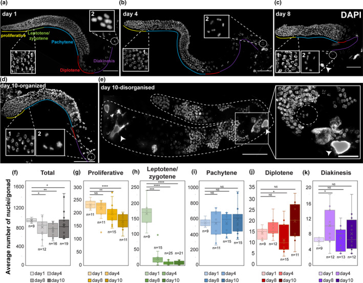
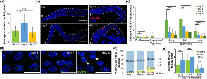
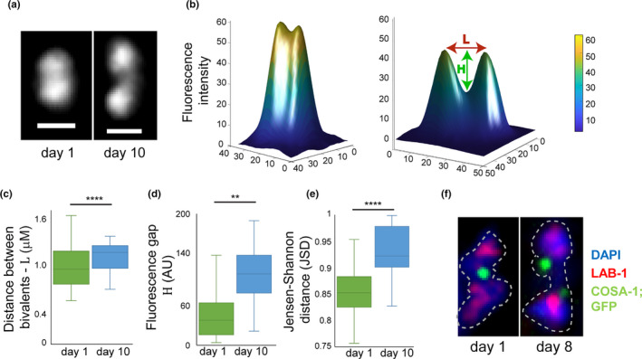
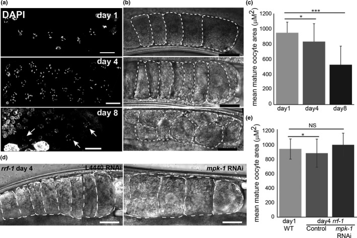
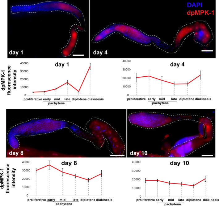
Oogenesis is one of the first processes to fail during aging. In women, most oocytes cannot successfully complete meiotic divisions already during the fourth decade of life. Studies of the nematode Caenorhabditis elegans have uncovered conserved genetic pathways that control lifespan, but our knowledge regarding reproductive aging in worms and humans is limited. Specifically, little is known about germline internal signals that dictate the oogonial biological clock. Here, we report a thorough characterization of the changes in the worm germline during aging. We found that shortly after ovulation halts, germline proliferation declines, while apoptosis continues, leading to a gradual reduction in germ cell numbers. In late aging stages, we observed that meiotic progression is disturbed and crossover designation and DNA double-strand break repair decrease. In addition, we detected a decline in the quality of mature oocytes during aging, as reflected by decreasing size and elongation of interhomolog distance, a phenotype also observed in human oocytes. Many of these altered processes were previously attributed to MAPK signaling variations in young worms. In support of this, we observed changes in activation dynamics of MPK-1 during aging. We therefore tested the hypothesis that MAPK controls oocyte quality in aged worms using both genetic and pharmacological tools. We found that in mutants with high levels of activated MPK-1, oocyte quality deteriorates more rapidly than in wild-type worms, whereas reduction of MPK-1 levels enhances quality. Thus, our data suggest that MAPK signaling controls germline aging and could be used to attenuate the rate of oogenesis quality decline.
Keywords: C. elegans; MAPK; aging; fertility; meiosis; oocyte; oogenesis.
© 2021 The Authors. Aging Cell published by Anatomical Society and John Wiley & Sons Ltd.
Conflict of interest statement
The authors declare that they have no conflict of interest.
Figures
Similar articles
-
Coordination of Recombination with Meiotic Progression in the Caenorhabditis elegans Germline by KIN-18, a TAO Kinase That Regulates the Timing of MPK-1 Signaling.Genetics. 2016 Jan;202(1):45-59. doi: 10.1534/genetics.115.177295. Epub 2015 Oct 28. Genetics. 2016. PMID: 26510792 Free PMC article.
-
Progression of Meiosis Is Coordinated by the Level and Location of MAPK Activation Via OGR-2 in Caenorhabditis elegans.Genetics. 2019 May;212(1):213-229. doi: 10.1534/genetics.119.302080. Epub 2019 Mar 13. Genetics. 2019. PMID: 30867196 Free PMC article.
-
Multiple functions and dynamic activation of MPK-1 extracellular signal-regulated kinase signaling in Caenorhabditis elegans germline development.Genetics. 2007 Dec;177(4):2039-62. doi: 10.1534/genetics.107.081356. Genetics. 2007. PMID: 18073423 Free PMC article.
-
Start me up: cell signaling and the journey from oocyte to embryo in C. elegans.Dev Dyn. 2006 Mar;235(3):571-85. doi: 10.1002/dvdy.20662. Dev Dyn. 2006. PMID: 16372336 Review.
-
Oogenesis in Caenorhabditis elegans.Sex Dev. 2023;17(2-3):73-83. doi: 10.1159/000531019. Epub 2023 May 11. Sex Dev. 2023. PMID: 37232019 Free PMC article. Review.
Cited by
-
3,3'-Diindolylmethane Supplementation Maintains Oocyte Quality by Reducing Oxidative Stress and CEP-1/p53-Mediated Regulation of Germ Cells in a Reproductively Aged Caenorhabditis elegans Model.Antioxidants (Basel). 2022 May 11;11(5):950. doi: 10.3390/antiox11050950. Antioxidants (Basel). 2022. PMID: 35624814 Free PMC article.
-
The roles of TGFβ and serotonin signaling in regulating proliferation of oocyte precursors and germline aging.bioRxiv [Preprint]. 2024 May 9:2024.05.08.593208. doi: 10.1101/2024.05.08.593208. bioRxiv. 2024. PMID: 38766220 Free PMC article. Preprint.
-
NuRD chromatin remodeling is required to repair exogenous DSBs in the Caenorhabditis elegans germline.bioRxiv [Preprint]. 2024 Sep 15:2024.09.14.613027. doi: 10.1101/2024.09.14.613027. bioRxiv. 2024. PMID: 39314477 Free PMC article. Preprint.
-
Aging and sperm signals alter DNA break formation and repair in the C. elegans germline.PLoS Genet. 2022 Nov 7;18(11):e1010282. doi: 10.1371/journal.pgen.1010282. eCollection 2022 Nov. PLoS Genet. 2022. PMID: 36342909 Free PMC article.
-
Reproductive Aging in Caenorhabditis elegans: From Molecules to Ecology.Front Cell Dev Biol. 2021 Sep 16;9:718522. doi: 10.3389/fcell.2021.718522. eCollection 2021. Front Cell Dev Biol. 2021. PMID: 34604218 Free PMC article. Review.
References
-
- Alpi, A. , Pasierbek, P. , Gartner, A. , & Loidl, J. (2003). Genetic and cytological characterization of the recombination protein RAD‐51 in Caenorhabditis elegans . Chromosoma, 112, 6–16. - PubMed
Publication types
MeSH terms
Substances
LinkOut - more resources
Full Text Sources
