. 2021 Jun 29;12(3):e0020221.
doi: 10.1128/mBio.00202-21. Epub 2021 Jun 1.

Potent Killing of Pseudomonas aeruginosa by an Antibody-Antibiotic Conjugate

Affiliations

Potent Killing of Pseudomonas aeruginosa by an Antibody-Antibiotic Conjugate

Kimberly K Kajihara et al. mBio. .

Abstract

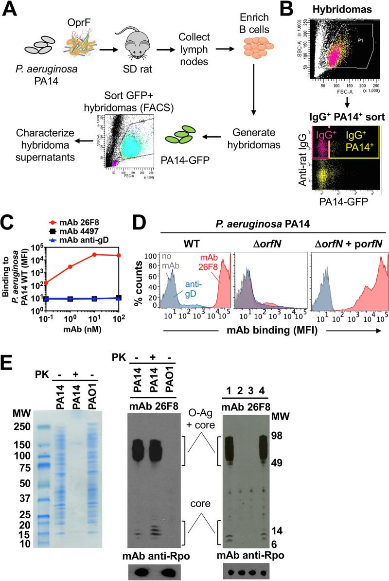
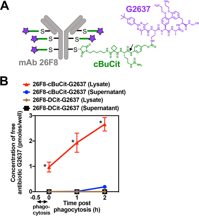
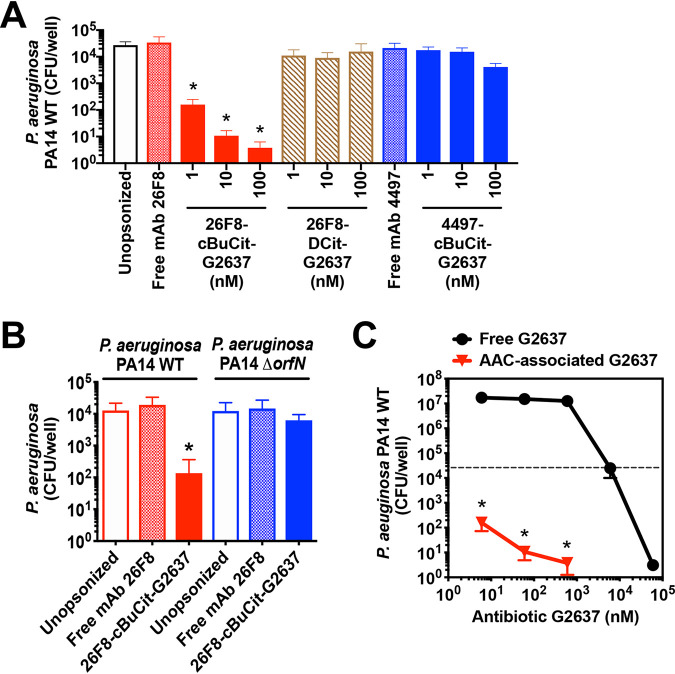
Pseudomonas aeruginosa causes life-threatening infections that are associated with antibiotic failure. Previously, we identified the antibiotic G2637, an analog of arylomycin, targeting bacterial type I signal peptidase, which has moderate potency against P. aeruginosa. We hypothesized that an antibody-antibiotic conjugate (AAC) could increase its activity by colocalizing P. aeruginosa bacteria with high local concentrations of G2637 antibiotic in the intracellular environment of phagocytes. Using a novel technology of screening for hybridomas recognizing intact bacteria, we identified monoclonal antibody 26F8, which binds to lipopolysaccharide O antigen on the surface of P. aeruginosa bacteria. This antibody was engineered to contain 6 cysteines and was conjugated to the G2637 antibiotic via a lysosomal cathepsin-cleavable linker, yielding a drug-to-antibody ratio of approximately 6. The resulting AAC delivered a high intracellular concentration of free G2637 upon phagocytosis of AAC-bound P. aeruginosa by macrophages, and potently cleared viable P. aeruginosa bacteria intracellularly. The molar concentration of AAC-associated G2637 antibiotic that resulted in elimination of bacteria inside macrophages was approximately 2 orders of magnitude lower than the concentration of free G2637 required to eliminate extracellular bacteria. This study demonstrates that an anti-P. aeruginosa AAC can locally concentrate antibiotic and kill P. aeruginosa inside phagocytes, providing additional therapeutic options for antibiotics that are moderately active or have an unfavorable pharmacokinetics or toxicity profile. IMPORTANCE Antibiotic treatment of life-threatening P. aeruginosa infections is associated with low clinical success, despite the availability of antibiotics that are active in standard microbiological in vitro assays, affirming the need for new therapeutic approaches. Antibiotics often fail in the preclinical stage due to insufficient efficacy against P. aeruginosa. One potential strategy is to enhance the local concentration of antibiotics with limited inherent anti-P. aeruginosa activity. This study presents proof of concept for an antibody-antibiotic conjugate, which releases a high local antibiotic concentration inside macrophages upon phagocytosis, resulting in potent intracellular killing of phagocytosed P. aeruginosa bacteria. This approach may provide new therapeutic options for antibiotics that are dose limited.

Keywords: Pseudomonas aeruginosa; antibiotics; antibody-antibiotic conjugate; macrophage.


Figures

FIG 1
FIG 1
Generation and characterization of MAb 26F8 recognizing LPS O antigen on P. aeruginosa bacteria. (A) Schematic of the immunization and sorting procedure. Rats were immunized with P. aeruginosa PA14 bacteria and boosted with OprF beta-barrel protein reconstituted in amphipols. Rat lymph nodes were harvested, and purified B cells were fused with Sp2ab cells to generate hybridomas, which were subjected to fluorescence-activated cell sorting (FACS) based on binding to GFP-labeled P. aeruginosa PA14. Supernatants were purified, and clones were selected based on positive binding to whole P. aeruginosa bacteria, as determined by FACS. (B) FACS sorting profile of rat hybridomas to select P. aeruginosa-binding antibodies. Rat hybridoma cells were incubated with fluorescent anti-rat IgG antibodies and with P. aeruginosa PA14 bacteria expressing GFP and were sorted from the IgG+ PA14+ gate (yellow; upper right quadrant). (C) Intact P. aeruginosa PA14 WT bacteria were incubated with MAbs and fluorescently labeled anti-human secondary antibodies, followed by determination of the mean fluorescence intensity (MFI; arbitrary units) by flow cytometry. Rat MAb 26F8, engineered with human Fc (red circles), showed dose-dependent, high-intensity binding to P. aeruginosa. Binding was hardly detectable for isotype-matched anti-S. aureus MAb 4497 (black squares) or for MAb against cytomegalovirus gD (anti-gD; blue triangles). (D) MAb 26F8 demonstrated high-intensity binding to intact P. aeruginosa PA14 WT bacteria (left). MAb 26F8 did not show binding to PA14 ΔorfN bacteria, which lack LPS O antigen (middle). Binding of MAb 26F8 to PA14 ΔorfN was restored by complementation with pUCP19-orfN (porfN) plasmid (right). Binding of MAb 26F8 to PA14 WT was represented by a shift in fluorescence of approximately 3 log compared to background fluorescence without antibody. Antibody binding was assessed by flow cytometry and was expressed as MFI. Red, MAb 26F8; blue, MAb anti-gD; gray, no MAb; MAbs were incubated at 10 nM. (E) (Left) Whole-cell lysates of P. aeruginosa PA14 WT or PAO1 WT were treated with or without proteinase K (PK), separated on SDS-PAGE gels, and stained with Coomassie. (Middle) Lysates of P. aeruginosa strains PA14 WT and PAO1 WT, treated with or without PK, were immunoblotted with MAb 26F8 (top) or, as a protein loading control, with MAb anti-RNA polymerase-α (Rpo) (bottom). (Right) Lysates of P. aeruginosa PA14 WT (lane 1), ΔorfN (lane 2), ΔorfN plus empty pUCP19 plasmid (lane 3), or ΔorfN plus pUCP19-orfN (lane 4) were immunoblotted with MAb 26F8 (top) or, as a loading control, with anti-Rpo MAb (bottom). MW, molecular weight marker.
FIG 2
FIG 2
Composition of the AAC molecule and delivery of high AAC-released intracellular concentrations of free G2637 antibiotic into macrophages. (A) Schematic of the 26F8-cBuCit-G2637 AAC molecule, composed of MAb 26F8 (gray), which recognizes the LPS O antigen of P. aeruginosa PA14, cathepsin-cleavable cBuCit linker (green), and the arylomycin analog antibiotic G2637 (purple), with a drug-to-antibody ratio of 6. The arrow indicates the cathepsin cleavage site. (B) Determination of the amount of free AAC-released antibiotic G2637 in cell lysates or supernatants after phagocytosis of AAC-preincubated P. aeruginosa PA14 WT by LC-MS/MS analysis. When PA14 WT bacteria were incubated with 26F8-cBuCit-G2637, free G2637 was detected in lysates immediately after 30 min of phagocytosis by macrophages and removal of extracellular bacteria (0 h); this value increased during 2 h of subsequent incubation following phagocytosis, suggesting continued intracellular cleavage of the linker. Free G2637 was hardly detectable in the extracellular cell supernatant, indicating prolonged intracellular retention of the free AAC-released antibiotic. The AAC 26F8-DCit-G2637, which contains the noncleavable DCit linker, did not release detectable intracellular G2637. Value are averages ± SD for technical triplicates; asterisks indicate significant differences (P < 0.01) from values for 26F8-DCit-G2637.
FIG 3
FIG 3
26F8-cBuCit-G2637 AAC induces potent intracellular killing of P. aeruginosa in macrophages. To determine anti-P. aeruginosa potency of AAC, P. aeruginosa bacteria were preincubated with AAC (1, 10, or 100 nM) or with free MAb (100 nM) and added to macrophages to induce phagocytosis. After addition of gentamicin to remove extracellular bacteria, macrophages were subsequently incubated for an additional 6 h at 37°C to enable bacterial killing, followed by macrophage lysis and CFU enumeration. (A) The 26F8-cBuCit-G2637 AAC (solid red bars) induced potent and dose-dependent intracellular killing of P. aeruginosa PA14 WT bacteria. In contrast, the viability of intracellular P. aeruginosa was hardly affected when bacteria were left unopsonized (empty bar) or preincubated with noncleavable 26F8-DCit-G2637 DAR6 AAC (hatched bars) or 4497-cBuCit-G2637 DAR6, which contains anti-S. aureus MAb 4497 (solid blue bars), or with free anti-P. aeruginosa MAb 26F8 (cross-hatched red bar) or free MAb 4497 (cross-hatched blue bar). (B) The 26F8-cBuCit-G2637 AAC promoted intracellular killing of P. aeruginosa PA14 WT (solid red bar) but not of the P. aeruginosa PA14 ΔorfN mutant (solid blue bar), which lacks the LPS O-antigen required for binding of MAb 26F8 (Fig. 1D and E). (A and B) Data are averages and SD for biological triplicates; asterisks indicate statistical significance (P < 0.05) compared to the CFU value of each condition immediately after phagocytosis (values are plotted in Fig. S3). (C) PA14 WT bacteria were preincubated with 26F8-cBuCit-G2637 AAC, followed by determination of killing 6 h postphagocytosis as for panel A (red triangles), or were incubated with free G2637 antibiotic for 6 h in the same medium without macrophages (black circles); viability of bacteria is expressed as CFU per well. 26F8-cBuCit-G2637-associated G2637 antibiotic required a molar concentration to kill P. aeruginosa PA14 WT approximately 2 orders of magnitude lower than that of free extracellular antibiotic G2637. The dashed line indicates input bacterial concentration at the start of the assay before the 6 h of incubation, which was in the same range for both conditions (approximately 2 × 104 to 5 × 104/ml). Value are averages ± SD for biological triplicates; asterisks indicate significant differences (P < 0.01) compared to free G2637 for each concentration.

References

    1. Thaden JT, Park LP, Maskarinec SA, Ruffin F, Fowler VG, van Duin D. 2017. Results from a 13-year prospective cohort study show increased mortality associated with bloodstream infections caused by Pseudomonas aeruginosa compared to other bacteria. Antimicrob Agents Chemother 61:e02671-16. - PMC - PubMed
    1. Planquette B, Timsit J-F, Misset BY, Schwebel C, Azoulay E, Adrie C, Vesin A, Jamali S, Zahar J-R, Allaouchiche B, Souweine B, Darmon M, Dumenil A-S, Goldgran-Toledano D, Mourvillier BH, Bédos J-P. 2013. Pseudomonas aeruginosa ventilator-associated pneumonia. Predictive factors of treatment failure. Am J Respir Crit Care Med 188:69–76. 10.1164/rccm.201210-1897OC. - DOI - PubMed
    1. Horcajada JP, Montero M, Oliver A, Sorlí L, Luque S, Gómez-Zorrilla S, Benito N, Grau S. 2019. Epidemiology and treatment of multidrug-resistant and extensively drug-resistant Pseudomonas aeruginosa infections. Clin Microbiol Rev 32:e00031-19. 10.1128/CMR.00031-19. - DOI - PMC - PubMed
    1. Cillóniz C, Gabarrús A, Ferrer M, de la Bellacasa JP, Rinaudo M, Mensa J, Niederman MS, Torres A. 2016. Community-acquired pneumonia due to multidrug- and non-multidrug-resistant Pseudomonas aeruginosa. Chest 150:415–425. 10.1016/j.chest.2016.03.042. - DOI - PubMed
    1. Zilberberg MD, Shorr AF. 2013. Prevalence of multidrug-resistant pseudomonas aeruginosa and carbapenem-resistant enterobacteriaceae among specimens from hospitalized patients with pneumonia and bloodstream infections in the United States from 2000 to 2009: drug resistance in pneumonia and BSI. J Hosp Med 8:559–563. 10.1002/jhm.2080. - DOI - PubMed

MeSH terms

Substances