. 2021 Jun 29;12(3):e0077121.
doi: 10.1128/mBio.00771-21. Epub 2021 Jun 1.

Obesity-Related Gut Microbiota Aggravates Alveolar Bone Destruction in Experimental Periodontitis through Elevation of Uric Acid

Affiliations

Obesity-Related Gut Microbiota Aggravates Alveolar Bone Destruction in Experimental Periodontitis through Elevation of Uric Acid

Keisuke Sato et al. mBio. .

Abstract

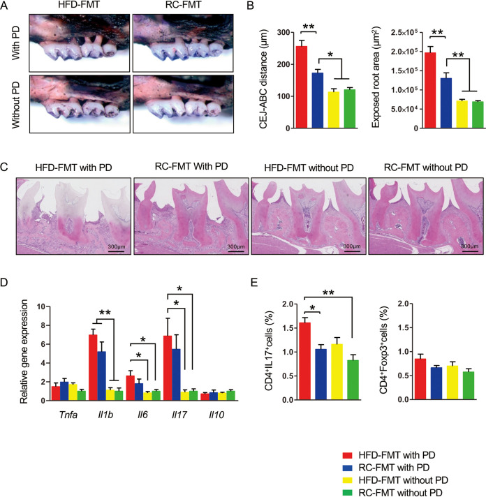
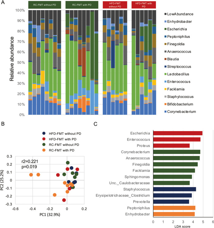
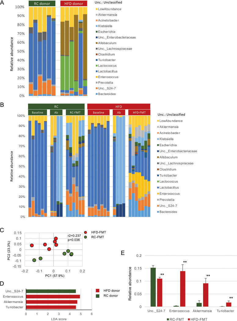
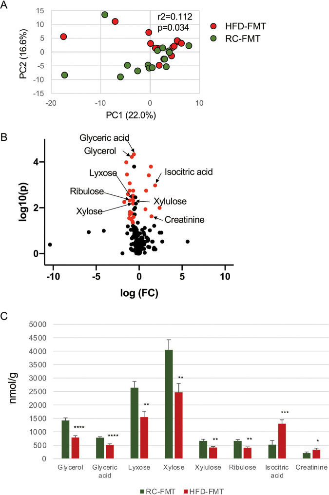
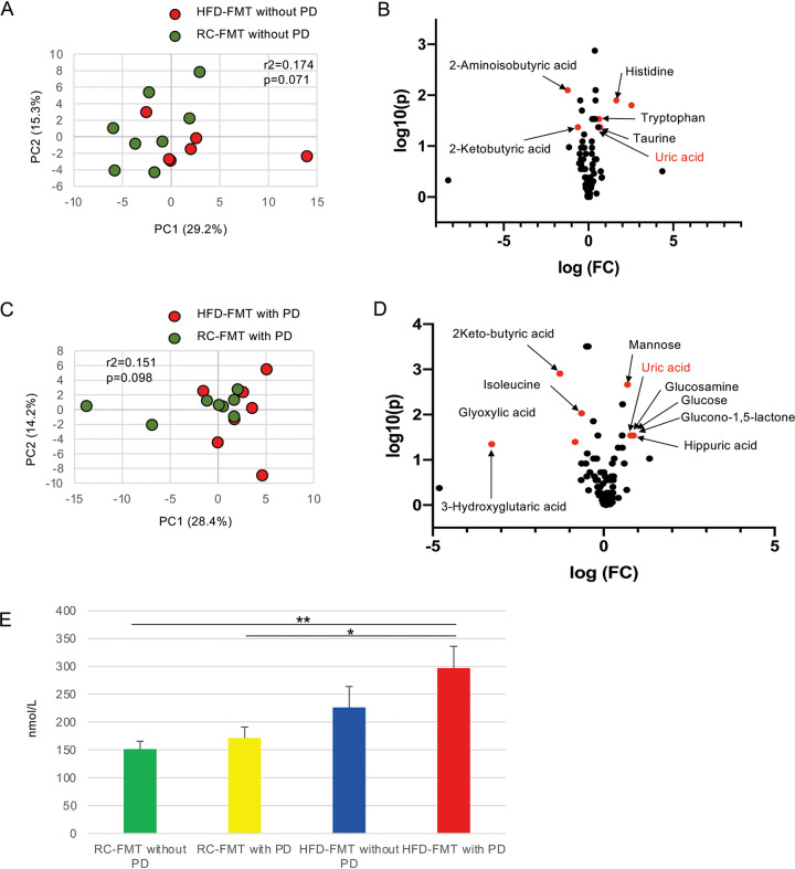
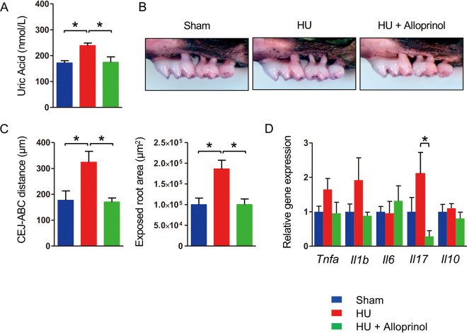
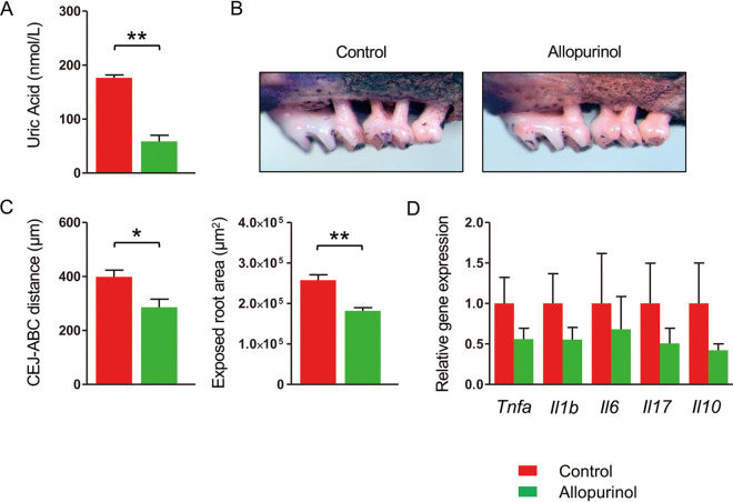
Obesity is a risk factor for periodontal disease (PD). Initiation and progression of PD are modulated by complex interactions between oral dysbiosis and host responses. Although obesity is associated with increased susceptibility to bacterial infection, the detailed mechanisms that connect obesity and susceptibility to PD remain elusive. Using fecal microbiota transplantation and a ligature-induced PD model, we demonstrated that gut dysbiosis-associated metabolites from high-fat diet (HFD)-fed mice worsen alveolar bone destruction. Fecal metabolomics revealed elevated purine degradation pathway activity in HFD-fed mice, and recipient mice had elevated levels of serum uric acid upon PD induction. Furthermore, PD induction caused more severe bone destruction in hyperuricemic than normouricemic mice, and the worsened bone destruction was completely abrogated by allopurinol, a xanthine oxidase inhibitor. Thus, obesity increases the risk of PD by increasing production of uric acid mediated by gut dysbiosis. IMPORTANCE Obesity is an epidemic health issue with a rapid increase worldwide. It increases the risk of various diseases, including periodontal disease, an oral chronic infectious disease. Although obesity increases susceptibility to bacterial infection, the precise biological mechanisms that link obesity and susceptibility to periodontal disease remain elusive. Using fecal microbial transplantation, experimental periodontitis, and metabolomics, our study demonstrates uric acid as a causative substance for greater aggravation of alveolar bone destruction in obesity-related periodontal disease. Gut microbiota from obese mice upregulated the purine degradation pathway, and the resulting elevation of serum uric acid promoted alveolar bone destruction. The effect of uric acid was confirmed by administration of allopurinol, an inhibitor of xanthine oxidase. Overall, our study provides new insights into the pathogenic mechanisms of obesity-associated periodontal disease and the development of new therapeutic options for the disease.

Keywords: gut microbiome; metabolomics; obesity; periodontal disease; uric acid.


Figures

FIG 1
FIG 1
Fecal microbial transplantation (FMT) affects the response of periodontal tissue in ligature-induced periodontitis (PD). Male C57BL/6N mice received FMT from regular chow (RC)- or high-fat diet (HFD)-fed mice and were either subjected to or not subjected to experimental periodontitis by ligature placement on the maxillary second molar (n = 5/group). (A) Effects of ligature placement on alveolar bone resorption in mice that received FMT from RC-fed (RC-FMT) or HFD-fed (HFD-FMT) mice. Representative photographs obtained after soft tissue removal are shown. (B) The distance between the cementoenamel junction and alveolar bone crest and the exposed tooth root area of the mesial root of the maxillary second molar was measured under a stereoscopic microscope. Red, HFD-FMT with PD; blue, RC-FMT with PD; yellow, HFD-FMT without PD; green, RC-FMT without PD. (C) Histological findings of gingival tissues of ligated and untreated mice. Sections were stained with hematoxylin and eosin. Representative photographs are shown. (D) Relative gene expression levels in the gingiva of each experimental group. The relative quantity of mRNA was normalized to that of glyceraldehyde-3-phosphate dehydrogenase mRNA. (E) Lymphocyte fractions were obtained from submandibular lymph nodes. Cells were stimulated with PMA and ionomycin at a concentration of 1 × 106/ml. The cells were stained with anti-CD4 and anti-IL-17 antibodies, and 1 × 104 cells were analyzed by flow cytometry. The percentages of CD4+IL-17+ and CD4+FoxP3+ cells were compared. Data are expressed as the mean ± standard error of the mean (SEM). *, P < 0.05; **, P < 0.01; one-way analysis of variance (ANOVA) with Bonferroni’s correction for multiple comparisons.
FIG 2
FIG 2
The oral microbiome is affected by ligature-induced periodontitis (PD) and FMT. FMT-treated mice with or without ligature-induced PD (referred to as RC-FMT without PD [n = 8], RC-FMT with PD [n = 8], HFD-FMT without PD [n = 6], and HFD-FMT with PD [n = 6]) were used in this experiment. (A) Relative abundance of oral bacterial taxa at the genus level in each experimental group. (B) Principal coordinate analysis (PCoA) score plot of the oral microbiota profiles of the four groups using unweighted UniFrac distance (ANOSIM). (C) Linear discriminant analysis (LDA) scores from LEfSe analysis. Enriched taxa in RC-FMT mice without PD, RC-FMT with PD, HFD-FMT without PD, and HFD-FMT with PD are indicated in green, yellow, blue, and red, respectively.
FIG 3
FIG 3
Different diets and FMT from mice fed different diets affect the gut microbiota. Feces obtained before (baseline) and after antibiotic treatment (Ab), after FMT (RC-FMT and HFD-FMT), and from donor mice (RC donor and HFD donor) were used in this experiment. (A) Relative abundance of gut bacterial taxa at the genus level in RC-fed (RC donor) and HFD-fed (HFD donor) mice. (B) Relative abundance of gut bacterial taxa at the genus level at baseline, after antibiotic treatment, and after fecal transplantation from RC-fed and HFD-fed mice. (C) Principal coordinate analysis (PCoA) score plot of the gut microbiota profiles of RC-FMT (n = 6) and HFD-FMT (n = 6) mice using unweighted UniFrac distance (ANOSIM). FMT from RC-fed and HFD-fed mice significantly affected recipient gut microbiota. (D) LDA scores from LEfSe analysis of fecal gut microbiota RC- and HFD-fed mice. (E) Pairwise comparisons of significantly changed bacterial taxa between RC-FMT and HFD-FMT mice. **, P < 0.01; Mann-Whitney U-test.
FIG 4
FIG 4
Difference in the gut metabolomic profile induced by the source of FMT. (A) Principal-component analysis (PCA) of the gut metabolomic profiles in RC-FMT (n = 16) and HFD-FMT (n = 13) mice. FMT from RC- and HFD-fed mice significantly affected recipient gut metabolomic profiles. (B) Volcano plot showing individual metabolites of RC-FMT and HFD-FMT mice. Red plots represent significantly different metabolites (fold change of >1.5 and P < 0.05). (C) Pairwise comparisons of significantly changed metabolites between RC-FMT and HFD-FMT mice. *, P < 0.05; **, P < 0.01; ***, P < 0.001; ****, P < 0.0001; Mann-Whitney U-test.
FIG 5
FIG 5
Difference in the serum metabolomic profile indued by the source of FMT and induction of periodontitis (PD). (A) PCA of the serum metabolomic profiles of RC-FMT without PD (n = 8) and HFD-FMT without PD (n = 6) mice. (B) Volcano plot showing individual serum metabolites in RC-FMT without PD and HFD-FMT without PD mice. (C) PCA of the serum metabolomic profiles of RC-FMT with PD (n = 8) and HFD-FMT with PD (n = 7) mice. (D) Volcano plot showing individual serum metabolites in RC-FMT with PD and HFD-FMT with PD mice. Red plots represent significantly different metabolites (fold change of >1.5 and P < 0.05). (E) Comparison of serum levels of uric acid after FMT and with or without induction of PD. *, P < 0.05; **, P < 0.01; Mann-Whitney U-test or one-way ANOVA with Bonferroni’s correction for multiple comparisons.
FIG 6
FIG 6
Hyperuricemia (HU) aggravates ligature-induced periodontitis. Three days after ligature placement, C57BL/6N mice were administered PBS, uric acid (125 mg/kg), or uric acid plus allopurinol (5 mg/kg) once a day for 4 days. PBS and uric acid were administered intraperitoneally, and allopurinol was administered via gastric gavage (n = 4 for uric acid and uric acid plus allopurinol, n = 3 for PBS). (A) Serum uric acid level in each group. Data are expressed as the mean ± SEM. (B) Effects of hyperuricemia on alveolar bone resorption. Representative images obtained after soft tissue removal are shown. (C) The distance between the cementoenamel junction and alveolar bone crest and the exposed tooth root area of the mesial root of the maxillary second molar was measured under a stereoscopic microscope. (D) Relative gene expression levels in the gingiva of each experimental group. The relative quantity of mRNA was normalized to that of glyceraldehyde-3-phosphate dehydrogenase mRNA. *, P < 0.05; one-way ANOVA with Bonferroni’s correction for multiple comparisons.
FIG 7
FIG 7
Allopurinol suppresses experimental periodontitis (PD) in HFD-FMT mice. Mice that received FMT from high-fat diet-fed mice were subjected to ligature-induced PD for 1 week. The mice were administered either allopurinol (5 mg/kg) (n = 6) or PBS (n = 6) every other day during the PD period. (A) Serum uric acid level in each group. (B) Effects of allopurinol on alveolar bone resorption. Representative images obtained after soft tissue removal are shown. (C) The distances between the cementoenamel junction and alveolar bone crest and the exposed tooth root area of the mesial root of the maxillary second molar were measured under a stereoscopic microscope. (D) Relative gene expression levels in the gingiva of each experimental group. The relative quantity of mRNA was normalized to that of glyceraldehyde-3-phosphate dehydrogenase mRNA. Data are expressed as the mean ± SEM. *, P < 0.05; **, P < 0.01; Mann-Whitney U-test.

References

    1. Suvan JE, Finer N, D’Aiuto F. 2018. Periodontal complications with obesity. Periodontol 2000 78:98–128. doi: 10.1111/prd.12239. - DOI - PubMed
    1. Hajishengallis G, Darveau RP, Curtis MA. 2012. The keystone-pathogen hypothesis. Nat Rev Microbiol 10:717–725. doi: 10.1038/nrmicro2873. - DOI - PMC - PubMed
    1. Gilbert JA, Blaser MJ, Caporaso JG, Jansson JK, Lynch SV, Knight R. 2018. Current understanding of the human microbiome. Nat Med 24:392–400. doi: 10.1038/nm.4517. - DOI - PMC - PubMed
    1. Costantini L, Molinari R, Farinon B, Merendino N. 2017. Impact of omega-3 fatty acids on the gut microbiota. Int J Mol Sci 18:2645. doi: 10.3390/ijms18122645. - DOI - PMC - PubMed
    1. Kesavalu L, Bakthavatchalu V, Rahman MM, Su J, Raghu B, Dawson D, Fernandes G, Ebersole JL. 2007. Omega-3 fatty acid regulates inflammatory cytokine/mediator messenger RNA expression in Porphyromonas gingivalis-induced experimental periodontal disease. Oral Microbiol Immunol 22:232–239. doi: 10.1111/j.1399-302X.2007.00346.x. - DOI - PubMed

Publication types