. 2021 Jun 29;12(3):e0326920.
doi: 10.1128/mBio.03269-20. Epub 2021 Jun 1.

GP96 Drives Exacerbation of Secondary Bacterial Pneumonia following Influenza A Virus Infection

Affiliations

GP96 Drives Exacerbation of Secondary Bacterial Pneumonia following Influenza A Virus Infection

Tomoko Sumitomo et al. mBio. .

Abstract

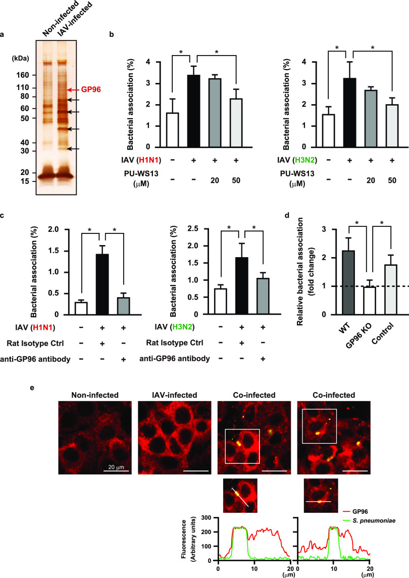
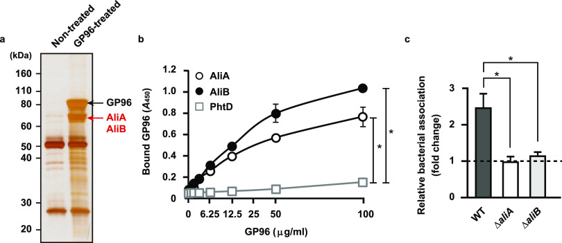
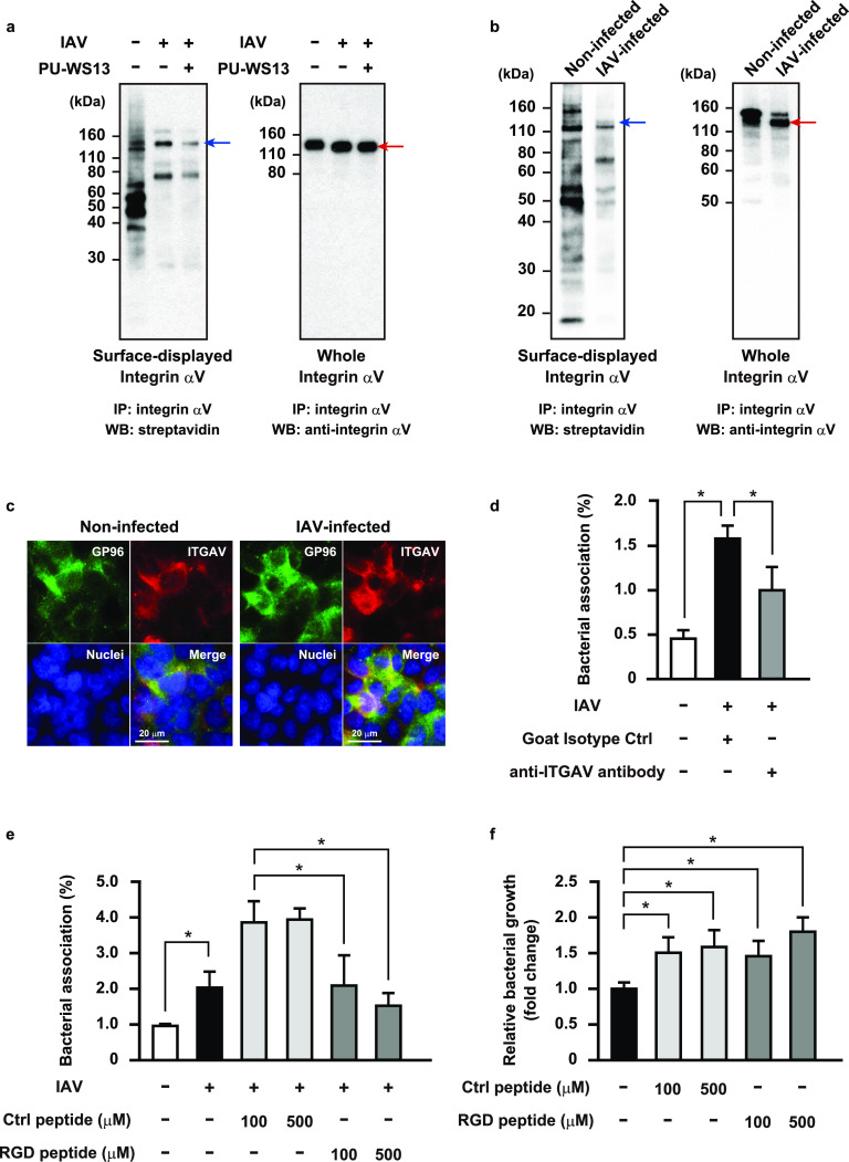
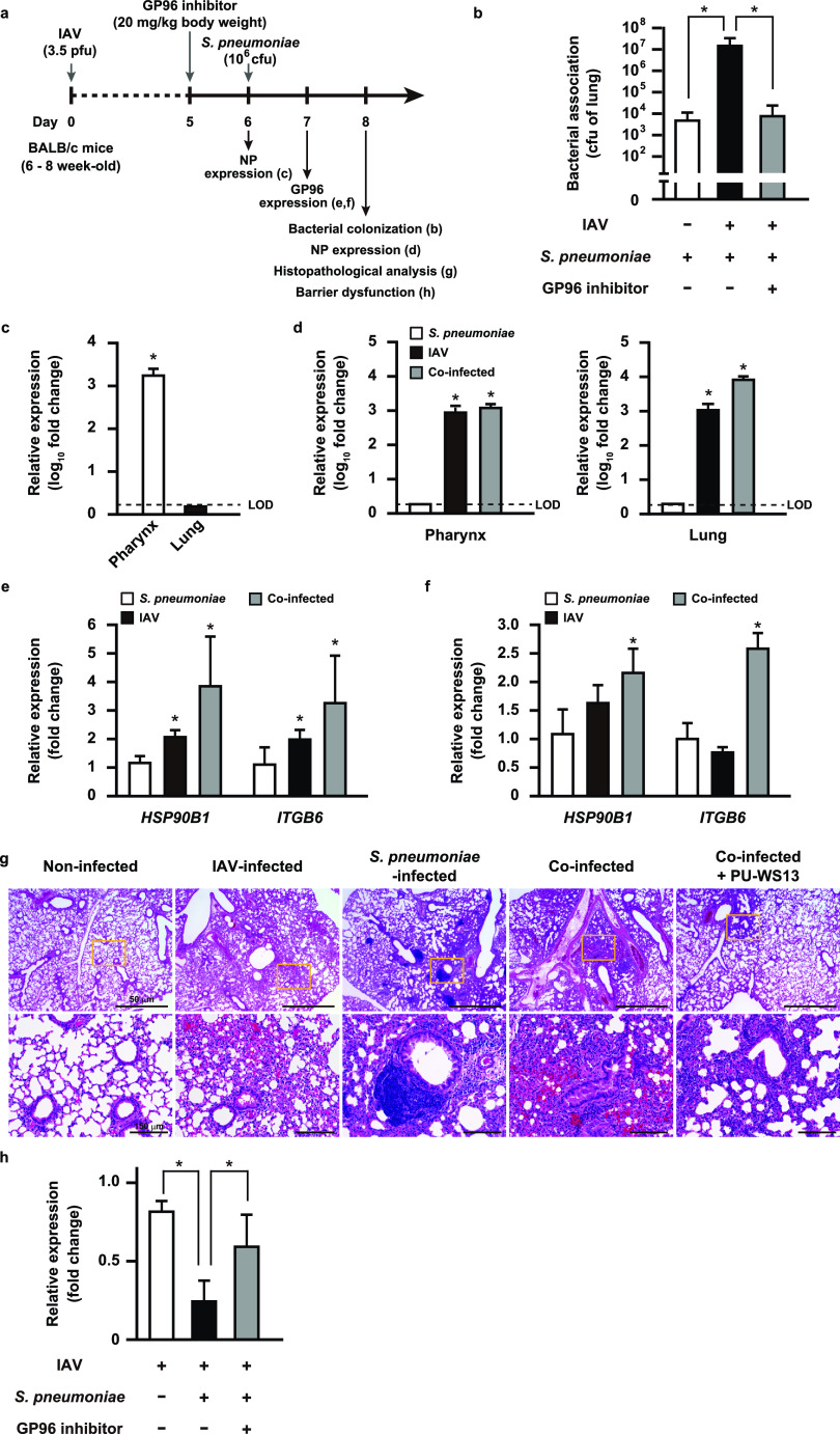
Influenza A virus (IAV) infection predisposes the host to secondary bacterial pneumonia, known as a major cause of morbidity and mortality during influenza virus epidemics. Analysis of interactions between IAV-infected human epithelial cells and Streptococcus pneumoniae revealed that infected cells ectopically exhibited the endoplasmic reticulum chaperone glycoprotein 96 (GP96) on the surface. Importantly, efficient pneumococcal adherence to epithelial cells was imparted by interactions with extracellular GP96 and integrin αV, with the surface expression mediated by GP96 chaperone activity. Furthermore, abrogation of adherence was gained by chemical inhibition or genetic knockout of GP96 as well as addition of RGD peptide, an inhibitor of integrin-ligand interactions. Direct binding of extracellular GP96 and pneumococci was shown to be mediated by pneumococcal oligopeptide permease components. Additionally, IAV infection induced activation of calpains and Snail1, which are responsible for degradation and transcriptional repression of junctional proteins in the host, respectively, indicating increased bacterial translocation across the epithelial barrier. Notably, treatment of IAV-infected mice with the GP96 inhibitor enhanced pneumococcal clearance from lung tissues and ameliorated lung pathology. Taken together, the present findings indicate a viral-bacterial synergy in relation to disease progression and suggest a paradigm for developing novel therapeutic strategies tailored to inhibit pneumococcal colonization in an IAV-infected respiratory tract. IMPORTANCE Secondary bacterial pneumonia following an influenza A virus (IAV) infection is a major cause of morbidity and mortality. Although it is generally accepted that preceding IAV infection leads to increased susceptibility to secondary bacterial infection, details regarding the pathogenic mechanism during the early stage of superinfection remain elusive. Here, we focused on the interaction of IAV-infected cells and Streptococcus pneumoniae, which revealed that human epithelial cells infected with IAV exhibit a cell surface display of GP96, an endoplasmic reticulum chaperon. Notably, extracellular GP96 was shown to impart efficient adherence for secondary infection by S. pneumoniae, and GP96 inhibition ameliorated lung pathology of superinfected mice, indicating it to be a useful target for development of therapeutic strategies for patients with superinfection.

Keywords: Streptococcus pneumoniae; influenza virus; pneumonia; superinfection.


Figures

FIG 1
FIG 1
IAV infection-induced surface display of GP96 promotes pneumococcal adherence. (a) A549 cells were infected with 106 PFU of IAV for 36 h and then treated with a membrane-impermeable biotinylation regent. Cell surface proteins were obtained using streptavidin beads and then subjected to SDS-PAGE and silver staining. Arrows indicate bands of upregulated surface proteins after IAV infection. Data shown are representative of at least three independent experiments. (b and c) A549 cells were infected with IAV (H1N1 or H3N2) for 1 h. Following washing steps, they were incubated for 36 h in the presence of PU-WS13 (b) or an antibody against GP96 (c). Next, IAV-infected cells were coinfected with S. pneumoniae D39 strain at a multiplicity of infection (MOI) of 5. At 2 h after infection, cells were lysed and cell-associated bacteria were recovered. Bacterial adherence rate was calculated as percentage of the inoculum. All experiments were performed in sextuplets with three technical repeats. Values are shown as the means ± standard deviations (SDs) from six wells from a representative experiment. (d) Effect of GP96 knockout (KO) on pneumococcal adherence. Bacterial association with IAV-infected cells was normalized to that with noninfected cells. For control cells with no mutation of the GP96 gene, cells cultured in the same manner as the GP96 knockout cells during selection were utilized. All experiments were performed in sextuplets with three technical repeats. Data are shown as relative bacterial association normalized to that with noninfected cells. Values are shown the means ± SDs from six wells from a representative experiment. *, P < 0.01 (b to d). (e) A549 cells were infected with IAV followed by S. pneumoniae infection. GP96 was labeled with anti-GP96 and Alexa Fluor 594-conjugated antibodies (shown as red in images), while S. pneumoniae was labeled with anti-serotype 2 capsule and Alexa Fluor 488-conjugated antibodies (shown as green in images). Images were analyzed using a confocal laser scanning microscope. Boxed area is magnified and shown in the lower panels. Graphs below the magnified images show fluorescence intensity profiles from a line crossing through the image of a coinfected cell. Coincidence of fluorescence intensity peaks, both red and green, indicates colocalization of S. pneumoniae with GP96. Data shown are representative of at least three separate experiments.
FIG 2
FIG 2
S. pneumoniae adheres to alveolar epithelial cells through interaction of pneumococcal surface proteins with GP96. (a) Proteins bound to GP96 were immunoprecipitated from pneumococcal cell wall fractions and then subjected to SDS-PAGE and silver staining. Data shown are representative of at least three separate experiments. (b) AliA, AliB, and PhtD, bacterial surface proteins, were immobilized on microtiter plates and then increasing amounts of GP96 were added. Bound GP96 was detected using an anti-GP96 antibody. All experiments were performed in sextuplets with three technical repeats. Values are shown as the means ± SDs from six wells from a representative experiment. *, P < 0.01. (c) Effects of deletion of aliA and aliB on pneumococcal adherence. Bacterial association with IAV-infected cells was normalized to that with noninfected cells. All experiments were performed in sextuplets with three technical repeats. Values are shown as the means ± SDs from six wells from a representative experiment. *, P < 0.01.
FIG 3
FIG 3
GP96-dependent surface display of integrin αV associated with enhanced pneumococcal adherence following IAV infection. A549 cells (a) or GP96 knockout cells (b) were infected with IAV for 36 h in the presence or absence of PU-WS13 and then treated with a membrane-impermeable biotinylation reagent. Immunoprecipitation of cell lysates containing biotinylated surface proteins was performed using an antibody against integrin αV. Surface-displayed and whole-cell integrin αV was detected using streptavidin and an antibody against integrin αV, respectively. Red arrows indicate integrin αV bands. Blue arrows indicate predicted molecular weight of surface-displayed integrin αV. IP, immunoprecipitation; WB, western blotting. Data shown are representative of at least three separate experiments. (c) A549 cells were infected with IAV for 1 h. After transferring to fresh medium, incubation was continued for 36 h. GP96 was labeled with anti-GP96 and Alexa Fluor 488-conjugated antibodies, while integrin αV was labeled with anti-integrin αV and Alexa Fluor 594-conjugated antibodies. DAPI was used to stain DNA in the nucleus. Data shown are representative of at least three independent experiments. (d and e) A549 cells were infected with IAV for 36 h. Following washing steps, they were incubated with an antibody against integrin αV (d) or RGD peptide (e) for 1 h, and then IAV-infected cells were coinfected with an S. pneumoniae strain at an MOI of 5. At 2 h after initiating infection, cells were lysed and cell-associated bacteria were recovered. Bacterial adherence rate was calculated as percentage of inoculum. All experiments were performed in sextuplets with three technical repeats. Values are shown as the means ± SDs from six wells from a representative experiment. (f) S. pneumoniae organisms were grown in cell culture medium containing the control or RGD peptide for 2 h, and then viable bacteria were recovered. Bacterial growth rate was calculated as percentage of inoculum. All experiments were performed in sextuplets with three technical repeats. Values are shown as the means ± SDs from six wells from a representative experiment. *, P < 0.01.
FIG 4
FIG 4
Calpain and Snail1 related to destruction of alveolar epithelial barrier following IAV infection. (a) A549 cells were infected with IAV for 1 h. After transferring to fresh medium, incubation was continued for 36 h. GP96 was labeled with anti-GP96 and Alexa Fluor 594-conjugated antibodies, while calpains were labeled with anti-calpain 2 and Alexa Fluor 488-conjugated antibodies. DAPI was used to stain DNA in the nucleus. (b) Transcriptional levels of genes encoding junctional proteins and regulators in A549 cells infected with IAV were analyzed using real-time RT-PCR. The gapdh transcript served as an internal control. OCLN, occludin; CDH1, E-cadherin; CTNND1, p120-catenin; CTNNB1, β-catenin; CTNNA1, αE-catenin; SNAI1, Snail1; SNAI2, slug; HSP90B1, GP96; CAPN1, calpain 1; CAPN2, calpain 2. Data from three independent tests are presented, with values shown as the means ± SDs for expression ratios. Transcriptional levels of tested genes are presented as relative expression levels normalized to that of noninfected cells. *, P < 0.01 compared to noninfected group. (c) A549 cells were infected with IAV for 1 h and then incubated with fresh medium in the presence or absence of SB-431542. Following the washing steps, cells were infected with an S. pneumoniae strain for 7 h. Expressions of E-cadherin, p120-catenin, and Snail1 were detected in whole-cell lysates using western blot analysis. β-Actin served as a loading control. All data shown are representative of at least three separate experiments.
FIG 5
FIG 5
GP96 is a crucial factor for exacerbation of bacterial pneumonia following IAV infection. (a) Schematic showing experimental design. Mice were intranasally infected with IAV (day 0) followed by S. pneumoniae on day 6. In some experiments, PU-WS13, a GP96 inhibitor, was intratracheally administered. (b) Effect of PU-WS13 treatment on bacterial burden in lungs. Values shown represent the means ± SDs from quintuplet samples and are representative of at least three independent experiments. *, P < 0.01. Transcriptional levels of the NP gene encoding viral nucleoprotein in pharyngeal and lung tissues at 6 (c) or 8 (d) days after infection were analyzed using real-time RT-PCR. LOD, limit of detection. Transcriptional levels of HSP90B1 and ITGB6 genes encoding GP96 and integrin β6, respectively, in pharyngeal (e) and lung tissues (f) infected with IAV and S. pneumoniae were analyzed using real-time RT-PCR. The gapdh transcript served as an internal control. Values from three independent tests are presented as the means ± SDs for expression ratios (c to f). *, P < 0.01 compared to the noninfected group. (g) Lung tissues obtained from mice infected under various conditions were subjected to hematoxylin and eosin staining. Boxed areas are magnified and shown at the bottom. (h) Transcriptional levels of the E-cadherin gene in lung tissues infected under various conditions were analyzed by real-time RT-PCR. The gapdh transcript served as an internal control. Values from three independent tests are shown as the means ± SDs for expression ratios. Transcriptional levels are presented as relative expression levels normalized to that of noninfected tissues (c, d, e, f, and h). *, P < 0.01.

Similar articles

Cited by

References

    1. Zhang Q, Shi J, Deng G, Guo J, Zeng X, He X, Kong H, Gu C, Li X, Liu J, Wang G, Chen Y, Liu L, Liang L, Li Y, Fan J, Wang J, Li W, Guan L, Li Q, Yang H, Chen P, Jiang L, Guan Y, Xin X, Jiang Y, Tian G, Wang X, Qiao C, Li C, Bu Z, Chen H. 2013. H7N9 influenza viruses are transmissible in ferrets by respiratory droplet. Science 341:410–414. doi:10.1126/science.1240532. - DOI - PubMed
    1. Neumann G, Kawaoka Y. 2015. Transmission of influenza A viruses. Virology 479–480:234–246. doi:10.1016/j.virol.2015.03.009. - DOI - PMC - PubMed
    1. Liang L, Jiang L, Li J, Zhao Q, Wang J, He X, Huang S, Wang Q, Zhao Y, Wang G, Sun N, Deng G, Shi J, Tian G, Zeng X, Jiang Y, Liu L, Liu J, Chen P, Bu Z, Kawaoka Y, Chen H, Li C. 2019. Low polymerase activity attributed to PA drives the acquisition of the PB2 E627K mutation of H7N9 avian influenza virus in mammals. mBio 10:e01162-19. doi:10.1128/mBio.01162-19. - DOI - PMC - PubMed
    1. Morens DM, Taubenberger JK, Fauci AS. 2008. Predominant role of bacterial pneumonia as a cause of death in pandemic influenza: implications for pandemic influenza preparedness. J Infect Dis 198:962–970. doi:10.1086/591708. - DOI - PMC - PubMed
    1. Joseph C, Togawa Y, Shindo N. 2013. Bacterial and viral infections associated with influenza. Influenza Other Respir Viruses 7 (Suppl 2):105–113. doi:10.1111/irv.12089. - DOI - PMC - PubMed

Publication types

MeSH terms