. 2021 Jun 1;15(6):e0009473.
doi: 10.1371/journal.pntd.0009473. eCollection 2021 Jun.

Immune exhaustion in chronic Chagas disease: Pro-inflammatory and immunomodulatory action of IL-27 in vitro

Affiliations

Immune exhaustion in chronic Chagas disease: Pro-inflammatory and immunomodulatory action of IL-27 in vitro

María Ailén Natale et al. PLoS Negl Trop Dis. .

Abstract

In chronic Chagas disease, Trypanosoma cruzi-specific T-cell function decreases over time, and alterations in the homeostatic IL-7/IL-7R axis are evident, consistent with a process of immune exhaustion. IL-27 is an important immunoregulatory cytokine that shares T-cell signaling with IL-7 and other cytokines of the IL-12 family and might be involved in the transcriptional regulation of T-cell function. Here, we evaluated the expression and function of IL-27R in antigen-experienced T cells from subjects with chronic Chagas disease and assessed whether in vitro treatment with IL-27 and IL-7 might improve T. cruzi-specific polyfunctional T-cell responses. In vitro exposure of PBMCs to T. cruzi induced a downregulation of IL-27R in CD4+ T cells and an upregulation in CD8+ T cells in subjects without heart disease, while IL-27R expression remained unaltered in subjects with more severe clinical stages. The modulation of IL-27R was associated with functional signaling through STAT3 and STAT5 and induction of the downstream genes TBX21, EOMES and CXCL9 in response to IL-27. In vitro treatment of PBMCs with IL-27 and IL-7 improved monofunctional and polyfunctional Th1 responses, accompanied by the induction of IL-10 and Bcl-2 expression in subjects without heart disease but did not improve those in subjects with cardiomyopathy. Our findings support the process of desensitization of the IL-27/IL-27R pathway along with disease severity and that the pro-inflammatory and immunomodulatory mechanisms of IL-27 might be interconnected.


Conflict of interest statement

The authors have declared that no competing interests exist.

Figures

Fig 1
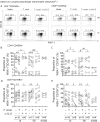
Fig 1. Differential expression of the IL-27R components in CD4+ and CD8+ antigen-experienced T cells in chronic T. cruzi-infected subjects with different degrees of cardiac dysfunction.
PBMCs from T. cruzi-infected and uninfected subjects (UI) were stained with the dye FV510 and the monoclonal antibodies for CD3, CD4, CD8, CD45RA, WSX-1 and CD130 and analyzed using flow cytometry. Each symbol represents the frequency of antigen-experienced CD45RAWSX-1+CD130+ (A, C) and CD45RAWSX-1CD130+ (B, D) CD4+ and CD8+ cells, relativized to the individual values of CD4+ and CD8+ for each subject. Medians are indicated by the horizontal lines; boxes indicate the 10–90 percentile range. *P < 0.05, ** P < 0.01 compared with UI by the Kruskal-Wallis test followed by Dunn’s test (A-B) or ANOVA followed by Bonferroni’s test (C-D), as appropriate.
Fig 2
Fig 2. Modulation of IL-27R components after in vitro infection of PBMCs with T. cruzi trypomastigotes.
The PBMCs of Chagas disease patients with no signs of heart disease (i.e., the G0 group) and with cardiomyopathy (i.e., the G1/G2 groups) and uninfected subjects (UI) were co-cultivated with T. cruzi trypomastigotes for 48 h. The cells were stained with the dye FV510 and the monoclonal antibodies for CD3, CD4, CD8, CD45RA, WSX1 and CD130 and analyzed using flow cytometry. Representative data of the pattern of WSX-1 and CD130 expression on CD4+CD45RA(A, left) or CD8+CD45RA(A, right) T cells of a G0 patient (upper panels) and an uninfected subject (lower panels). Data are shown as dot plots of basal IL-27R expression (media, left panels), IL-27R expression after coculture with T. cruzi trypomastigotes (T. cruzi, middle panels) and IL-27R expression after coculture with T. cruzi trypomastigotes and with the addition of anti-IL-2 neutralizing antibodies (T. cruzi + α-IL-2, right panels). The lower figures show the proportions of WSX-1+CD130+ (right quadrants) and WSX-1CD130+ (left quadrants). Each symbol represents the proportions of cells expressing both IL-27R components (WSX-1+CD130+) or with downmodulated WSX-1 chain (WSX-1CD130+) of each subject in CD45RAcells in the total CD4+ (B-C) and CD8+ (D-E) T cell compartments. * P < 0.05, ** P < 0.01, and *** P < 0.001 compared with unstimulated cultures using paired t-test. # P < 0.05, ## P < 0.01, and ### P < 0.001 indicate the differences in the ratio of T. cruzi-stimulated and unstimulated cultures in the presence or absence of neutralizing IL-2 antibodies in each clinical group using the Mann Whitney U test.
Fig 3
Fig 3. IL-27-induced phosphorylation of STAT1, STAT3, and STAT5 in vitro is reduced in chronic Chagas disease cardiomyopathy.
Each symbol represents STAT1 (A-B), STAT3 (C-D) and STAT5 (E-F) phosphorylation before and after stimulation with IL-27 of PBMCs in vitro (% pSTAT+) in CD4+ (A, C, E) and CD8+ (B, D, F) T cells as evaluated using flow cytometry. Each symbol represents the frequency of CD4+/CD8+pSTAT1+, pSTAT3+ and pSTAT5+ for individual subjects, relativized to the individual values of CD4+ and CD8+ for each subject. Comparisons of the frequency of CD4+ and CD8+ T cells expressing the corresponding pSTAT in IL-27-stimulated cell cultures and that in unstimulated cell-cultures in each group (shown as * P < 0.05; ** P < 0.01, *** < 0.001 and **** P < 0.0001) were performed by paired t test. Only P < 0.05, along with a median increase of at least 50% in pSTAT+ T cells, was considered statistically significant. Comparisons of the frequency of CD4+ and CD8+ T cells expressing the corresponding pSTAT in unstimulated cell-cultures (full gray symbols, shown as # P < 0.05; ## P < 0.01 and ### P < 0.001) among the clinical groups and the uninfected subjects (UI) were performed by a Kruskal-Wallis test followed by Dunn’s test for multiple comparisons. There is a positive trend in the percentages of constitutive CD4+ or CD8+ T cells expressing pSTAT1 (A, P = 0.025 slope (m) = 1.29 and B, P = 0.0019 m = 1.08); pSTAT3 (C, P = 0.025 m = 1.78 and D, P = 0.032 m = 1.83) and STAT5 (F, P = 0.0001 m = 1.08) as the clinical stage becomes more severe.
Fig 4
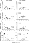
Fig 4. Expression of unmodulated IL-27R components associates with low STAT phosphorylation and IFN-γ production in response to T. cruzi antigens.
The Spearman test was applied to assess the correlation between the frequency of memory CD4+CD45RA(A, C and E) and CD8+CD45RA(B, D and F) T cells expressing both chains of the IL-27R and the IL-27-induced STAT phosphorylation (post-stimulation/pre-stimulation ratio) in CD4+ and CD8+ T cells, respectively; the frequency of CD4+ (G) and CD8+ (H) T cells expressing both chains of the IL-27R and the total number of IFN-γ-producing cells in response to T. cruzi antigens. Full symbols represent values from subjects without heart disease symptoms (i.e., the G0 group); open symbols represent values from subjects with cardiac dysfunction (i.e., the G1, G2 and G3 groups).
Fig 5
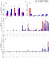
Fig 5. Polyfunctionality of T. cruzi-specific CD4+ T cells in patients with chronic Chagas disease with no sign of cardiomyopathy is improved after in vitro treatment with IL-27 and IL-7.
PBMCs of chronic T. cruzi-infected subjects with no signs of heart disease (n = 10) and subjects with heart disease (i.e., the G2 group, n = 4) were stimulated with a T. cruzi lysate preparation from the Brazil strain in the presence or absence of IL-27 (red bars) or IL-7 (blue bars) and analyzed using flow cytometry for the intracellular expression of TNF-α, MIP-1β, IL-2, IFN-γ and CD154 in CD4+ T cells. Each bar represents the frequency of total TNF-α-, MIP-1β-, IL-2-, IFN-γ- and CD154-T. cruzi-specific CD3+CD4+ T-cell responses (A and B). For the analysis of polyfunctional T-cell responses, the cytokine coexpression profiles with one (1+), two (2+), three (3+), four (4+) and five (5+) functions were determined using the Boolean gating function of FlowJo software. Each bar represents the total frequency of T. cruzi-specific (i.e., values obtained in cultures with only media were subtracted) CD3+CD4+ T-cell responses of each cytokine-producing subset relative to the individual values of CD4+ for each subject (C and D). * P < 0.05, ** P < 0.01, *** P < 0.001 compared with T. cruzi-specific T-cell responses without the addition of cytokine (white bars) according to a paired t test. Data are shown as the means and SD.
Fig 6
Fig 6. IL-10 production in response to IL-27 is impaired in Chagas disease patients with severe cardiomyopathy.
PBMCs of subjects with chronic Chagas disease with no signs of heart disease (i.e., the G0 group) or with myocardiopathy (i.e., the G2-G3 group) and of uninfected subjects (i.e., UI) were stimulated with 100 ng/mL IL-27 for 20 h and then evaluated for IL-10 production in CD4+ T cells using flow cytometry. Each point represents the frequency of CD4+IL-10+ T cells, relativized to the individual values of CD4+ and CD3+ for each subject. * P <0.05 compared with unstimulated cell culture according to a paired t test..# P < 0.05 between post-stimulation/pre-stimulation ratios of the indicated subject groups. Only P < 0.05, along with a median increase of at least 50% in IL-10+ T cells, was considered statistically significant.
Fig 7
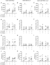
Fig 7. Bcl-2 expression is higher in IFN-γ- and IL-2-coproducing T cells in response to T. cruzi antigens after treatment with IL-27 and IL-7.
PBMCs of chronic T. cruzi-infected subjects with no signs of heart disease (n = 10) were stimulated with a T. cruzi lysate preparation from the Brazil strain in the presence or absence of IL-27 or IL-7. Flow cytometry analysis was applied to determine the expression of Bcl-2 (A-C), PD-1+ (D-F), and CD57+ (G-I) or the coexpression of PD-1 and CD57 (J-L) cells in IFN-γ- and IL-2-producing T cells. Each different symbol represents data for a specific patient. IL-27 or IL-7-stimulated and unstimulated samples are bound by lines. Gray symbols bound by dotted lines represent data of subjects in whom T. cruzi-specific cytokine-producing T cells in response to T. cruzi lysate were undetectable in the absence of IL-27 or IL-7 stimulation, which became detecs upon IL-27 or IL-7 stimulation. Data were analyzed by a paired t test. * P < 0.05; ** P < 0.01 compared with T. cruzi stimulated cell cultures without the addition of cytokines. Only P < 0.05, along with a median increase of at least 50% in cytokine-producing cells expressing Bcl-2, PD-1 or CD57, was considered statistically significant.

Similar articles

Cited by

References

    1. Bern C, Montgomery SP, Herwaldt BL, Rassi A, Marin-Neto JA, Dantas RO, et al.. Evaluation and treatment of Chagas disease in the United States: A systematic review. J Am Med Assoc. 2007;298: 2171–81. doi: 10.1001/jama.298.18.2171 - DOI - PubMed
    1. WHO. Fact sheet: Chagas disease (American trypanosomiasis) [Internet]. 2020 p. who.int/mediacentre/factsheets/fs340/en/. Available: who.int/mediacentre/factsheets/fs340/en/
    1. Tarleton RL. Chagas disease: A role for autoimmunity? Trends Parasitol. 2003;19: 447–451. doi: 10.1016/j.pt.2003.08.008 - DOI - PubMed
    1. Wherry EJ, Kurachi M. Molecular and cellular insights into T cell exhaustion. Nat Rev Immunol. 2015;15: 486–499. doi: 10.1038/nri3862 - DOI - PMC - PubMed
    1. Dutra WO, Menezes CAS, Villani FNA, da Costa GC, da Silveira ABM, Reis DDÁ, et al.. Cellular and genetic mechanisms involved in the generation of protective and pathogenic immune responses in human Chagas disease. Mem Inst Oswaldo Cruz. 2009;104: 208–218. doi: 10.1590/s0074-02762009000900027 - DOI - PMC - PubMed

Publication types

MeSH terms