. 2021 Jun 1;16(6):e0251873.
doi: 10.1371/journal.pone.0251873. eCollection 2021.

A novel approach to evaluate the effects of artificial bone focal lesion on the three-dimensional strain distributions within the vertebral body

Affiliations

A novel approach to evaluate the effects of artificial bone focal lesion on the three-dimensional strain distributions within the vertebral body

Marco Palanca et al. PLoS One. .

Abstract

The spine is the first site for incidence of bone metastasis. Thus, the vertebrae have a high potential risk of being weakened by metastatic tissues. The evaluation of strength of the bone affected by the presence of metastases is fundamental to assess the fracture risk. This work proposes a robust method to evaluate the variations of strain distributions due to artificial lesions within the vertebral body, based on in situ mechanical testing and digital volume correlation. Five porcine vertebrae were tested in compression up to 6500N inside a micro computed tomography scanner. For each specimen, images were acquired before and after the application of the load, before and after the introduction of the artificial lesions. Principal strains were computed within the bone by means of digital volume correlation (DVC). All intact specimens showed a consistent strain distribution, with peak minimum principal strain in the range -1.8% to -0.7% in the middle of the vertebra, demonstrating the robustness of the method. Similar distributions of strains were found for the intact vertebrae in the different regions. The artificial lesion generally doubled the strain in the middle portion of the specimen, probably due to stress concentrations close to the defect. In conclusion, a robust method to evaluate the redistribution of the strain due to artificial lesions within the vertebral body was developed and will be used in the future to improve current clinical assessment of fracture risk in metastatic spines.


Conflict of interest statement

The authors have declared that no competing interests exist.

Figures

Fig 1
Fig 1. Artificial lesions were reproduced in the anterior or lateral middle portion of the middle.
The position and volume of the lesions were identified in the microCT images.
Fig 2
Fig 2. Workflow of the image processing.
For each specimen, five scans were acquired to evaluate: the measurement uncertainties (Scan1-pre vs Scan1-crop), the strain field within the vertebral body of the intact vertebra (Scan1 vs Scan2), and the strain field within the vertebral body of the vertebra with artificial lesion (Scan3 vs Scan4). Pre-processing of the images, digital volume correlation (DVC) with the BoneDVC algorithm were used to measure the strain fields. A comparison between the two conditions was proposed.
Fig 3
Fig 3. Frequency plot for the εp1 and εp3 evaluated for each intact specimen for the entire vertebra, for the top, middle and bottom ROIs.
Fig 4
Fig 4
εp3 distributions in the intact vertebrae (left) and in the vertebrae with artificial lesions (middle). The volume of the lesion is reported close to the strain distributions for vertebrae with defects. On the right, rendering of the vertebra with the created artificial lesion and frequency plots for εp3 in the whole vertebral body with (orange) and without (blue) the lesions are reported. For the intact vertebrae, the larger strains localized in correspondence of the growth plates, both on the top and the bottom. For the vertebrae with the artificial lesions, the strain distribution was different and strain concentrations were visible around the lesion.
Fig 5
Fig 5
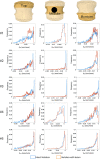
εp3 distributions were evaluated in the top (left column), middle (central column), and bottom (right column) longitudinal ROIs. The blue histograms refer to intact specimens, the orange ones to those with lesions.
Fig 6
Fig 6. Changes in the strain values at 6500N (exception for #2 and #3) for each subROI with and without the lesion.
The first column represents the scheme of the different subROIs above the central cross-sections before (green) and after (red) the lesion. The lesion mainly affected one single subROI, but may also involve adjacent subROI. Increase (red boxes) or decrease (blue boxes) of εp3 in the top (second column), middle (third column) and bottom (forth column) ROIs are reported on the right. Differences below the DVC measurement uncertainties are reported in grey.

Similar articles

Cited by

References

    1. Jacobs WB, Perrin RG. Evaluation and treatment of spinal metastases: an overview. FOC. 2001;11: 1–11. 10.3171/foc.2001.11.6.11 - DOI - PubMed
    1. Tomita K, Kawahara N, Kobayashi T, Yoshida A, Akamaru T. Surgical Strategy for Spinal Metastases. Spine. 2001;26: 298–306. 10.1097/00007632-200102010-00016 - DOI - PubMed
    1. Alkalay RN. Effet of the metastatic defect on the structural responde and failure process of human vertebrae: an experimental study. Clinical Biomechanics. 2015;30: 121–128. 10.1016/j.clinbiomech.2014.10.001 - DOI - PMC - PubMed
    1. Alkalay RN, Harrigan TP. Mechanical assessment of the effects of metastatic lytic defect on the structural response of human thoracolumbar spine: EFFECT OF CRITICAL LYTIC DEFECT. J Orthop Res. 2016;34: 1808–1819. 10.1002/jor.23154 - DOI - PubMed
    1. Palanca M, Barbanti-Bròdano G, Cristofolini L. The Size of Simulated Lytic Metastases Affects the Strain Distribution on the Anterior Surface of the Vertebra. Journal of Biomechanical Engineering. 2018;140: 111005. 10.1115/1.4040587 - DOI - PubMed