Review
. 2021 Nov 16;22(22):3109-3139.
doi: 10.1002/cbic.202100171. Epub 2021 Jun 23.

Lessons in Organic Fluorescent Probe Discovery

Affiliations
Review

Lessons in Organic Fluorescent Probe Discovery

Sachin B Wagh et al. Chembiochem. .

Abstract

Fluorescent probes have gained profound use in biotechnology, drug discovery, medical diagnostics, molecular and cell biology. The development of methods for the translation of fluorophores into fluorescent probes continues to be a robust field for medicinal chemists and chemical biologists, alike. Access to new experimental designs has enabled molecular diversification and led to the identification of new approaches to probe discovery. This review provides a synopsis of the recent lessons in modern fluorescent probe discovery.

Keywords: chemical biology; fluorescence; fluorophores; molecular probes; sensors.


Conflict of interest statement

Conflict of Interest

The authors declare no conflict of interest.

Figures

Figure 1.
Figure 1.
Exemplary fluorophores and fluorescent probes. a) Structure of matlaline, the presumptive first observed fluorescent compound. b) Fluorophore-conjugated phalloidin. Early work by Wieland in 1979 was one of the first studies on semi-synthetic preparation of a fluorescent derivative of phalloidin, where the sphere represents the position of the fluorescent tag. c) Structure of dansyl chloride and its amino acid conjugates.
Figure 2.
Figure 2.
Asian scorpion Mesobuthus martensii fluoresces under UVA light due to accumulated coumarins, such as 4-methylumbelliferone. The latter natural product has evolved to synthetic derivatives, such as Marina Blue, as a fluorescent reporter. Its phosphorylated derivative, DuFMUP is used as a probe for phosphatase activity. The image of the scorpion was used without editing and with permission from the authors under Creative Commons Attribution-NonCommercial-ShareAlike 3.0.[20]
Figure 3.
Figure 3.
Advancement from yellow curcumin to red rosocyanine and the NIR probe CRANAD-2. The color change from yellow (curcumin) to red (rosocyanine) could be explained by π→π* transition from oxygen to the empty orbital on boron. In addition, the replacement of phenolic hydroxyls in rosocyanine by N,N’-dimethyl groups in GRANAD-2 enabled a further red shift in emission.
Figure 4.
Figure 4.
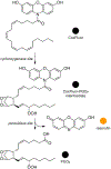
Proposed mechanism of COX-2-catalyzed resorufin production. The arachidonic acid tail serves as a substrate in a manner similar to the natural compound. Amide hydrolysis then releases the fluorophore resorufin and PGG2. Because COX-1 is unable to accommodate large groups at the substrate site, the bulky fluorophore imparts selectivity for COX-2 possessing a larger substrate pocket.
Figure 5.
Figure 5.
The structure-photophysical relationship (SPPR) in the Seoul-Fluor skeleton showing the intramolecular charge transfer (ICT) in the excited state (blue to red sites in the molecule). The structure of one example SF44,[32a] is provided as an illustration of the push-pull system wherein an electron-donating amine is conjugated to an electron-withdrawing acetyl group.
Figure 6.
Figure 6.
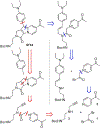
Retrosynthetic scheme for the Seoul-Fluor scaffold includes two synthetic routes that involve the assembly of three fragments. The first route (blue) begins by assembly of bromoacetyl bromide (1) and pyridines 2 to give substituted pyridines that react with reductive amination product from the coupling of cinnamaldehyde and a primary amine. The second route (red) begins by assembly of 1, pyridines 2, and propargyl amines 3 to generate an amide intermediate, which subsequently undergoes a 1,3-dipolar cycloaddition to produce the Seoul-Flour skeleton.
Figure 7.
Figure 7.
A schematic representation of DHP-based fluorophores. The 1,4-DHP fluorophore 4 has electron rich groups at 1-position and electron deficient groups at the 3- or 5-positions and generally exhibits blue fluorescence. The design of 1,2-dihydropyridine (DHP)-based fluorophore 5 includes arms for bioconjugation, chemical handle R1 and hydrolysable ester groups. The strategy of 1,2-DHP skeleton involves a push-pull system with different electron donor groups.
Figure 8.
Figure 8.
One-pot multicomponent synthesis of 1,2-DHP (5) derivatives from dienaminodioates (6), aldehydes (7) and in-situ generated hydrazones (8).
Figure 9.
Figure 9.
Structures of 1,2-dihydropyridines synthesized using a multicomponent one-pot reaction. The 6-position in N-benzylideneamine is the conjugation site. 1,2-DHPs 5ae emit in the long wavelength region of 500–600 nm.
Figure 10.
Figure 10.
Ribozyme-catalyzed Diels-Alder reaction between 9-hydroxymeth-ylanthracene (9) and N-pentylmaleimide (10).· The development of fluorescent probes for catalytic Diels-Alder reactions involves the optimization of such characteristics as sufficient solubility in an aqueous medium, diene-based reactivity identical to the substrate in the uncatalyzed Diels-Alder reactions, acceptance as a substrate by the ribozymes, the Diels-Alder reaction-based change in photophysical properties, as well as fluorescent changes brought about by binding to the ribozyme to observe and distinguish the key species of the catalytic cycle.
Figure 11.
Figure 11.
Design of fluorescent probe for the investigation of Diels-Alder reactions: substrate-analogue probes 13, 14 and product-analogue probe 12.
Figure 12.
Figure 12.
Schematic design of a Red-pHocas (pH activatable red fluorescent probe) for detecting the areas of bone acidification. This probe was used to demonstrate the direct involvement of osteoclast proton pumps in bone acidification.
Figure 13.
Figure 13.
A design strategy of dimethylaminoquinoline (DMAQ) fluorophore consisted of three domains, one polarization domain and two tuning domains. The retrosynthetic approach for DMAQ synthesis using cyclo-condensation of 3-(dimethylamino)aniline (15) and diethylmalonate followed by chlorination with phosphoryl chloride. Box: The structure of an exemplary DMAQ derivative 17.
Figure 14.
Figure 14.
Structures of fluorescein (18), carbofluorescein (19), a sulfonic acid analogue of carbofluorescein (20), and CarboVoltageFluor (21).
Figure 15.
Figure 15.
Retrosynthesis of CarboVoltageFluor (21). The assembly begins by synthesis of carbofluorescein precursor 24 using 3-chloro-4-methoxybenzoyl chloride (22) and 4-bromo-2-chloro-1-methoxybenzene (23) followed by the installation of the dimethylcarbon. Nucleophilic addition of an aryllithium species gave 26, which was followed by the Heck coupling of the phenylenevinylene ‘molecular wire’ in 22.
Figure 16.
Figure 16.
Boronic acid-based fluorescent probes for saccharide detection. Probes are designed by quenching the emission of fluorophores attached to an electron-donating molecular recognition element (photoinduced electron transfer) in order to achieve fluorophores that display enhanced fluorescence when bound to a saccharide.
Figure 17.
Figure 17.
Modular design of fluorescent sensory systems containing two boronic acid residues for detection of specific saccharides. When bound to a saccharide, the photoinduced electron transfer from amino group to the aryl unit within the probe is hindered thus resulting in a fluorescent response. Modular design allows for fine-tuning of selectivity and localization in biological systems.
Figure 18.
Figure 18.
Optical imaging of tumors through selective labeling of glycoproteins. Boronic acid-functionalized peptide-based fluorescent sensors are water-soluble and serve as biocompatible tools for in situ recognition and differentiation of cell surface carcinoma biomarkers in vivo.
Figure 19.
Figure 19.
Peptide-based Zn2+ fluorescent probes. a) Increase in fluorophore emission due to binding-induced peptide conformational regulation. Protein scaffold shields the fluorophore from solvent interactions. b) Structure of the Palm-ZP1 (34), which was used for localized Zn detection in live HeLa cells. The peptide contains a 3-residue polyproline helix, 2 sequential Asp residues and a C-terminal Lys to serve as the point of attachment for the Zn2+ probe.
Figure 20.
Figure 20.
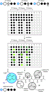
Analysis of proteolytic enzyme specificity through the release of conjugated fluorescent leaving group in the course of Positional Scanning Substrate Combinatorial Library method. Substrate specificity is assessed by measuring fluorescent response following the protease cleavage of a particular peptide bond.
Figure 21.
Figure 21.
Probe discovery process. Schematic representation of the probe discovery process illustrating how probes in libraries P1–P3 are prepared and screened to deliver an active probe 35.
Figure 22.
Figure 22.
Systematic screening of diversity-oriented fluorescent libraries. a) An example heat map plot of change in emission intensity after interaction of a probe with a target organized by probe. b) Selected fluorescent probes discovered by Chang and co-workers through unbiased screening of diversity-oriented fluorescent libraries.
Figure 23.
Figure 23.
Solid-supported diversity-oriented fluorescent probe libraries and array sensing. Synthesis of metal ion sensors supported on filter paper by in situ amination and carbodiimide coupling. Simultaneous response from multiple sensors can discriminate up to 12 metal ions. Newly discovered sensors can be utilized as unbound molecules or can be used as paper-based sensing devices.
Figure 24.
Figure 24.
Fingerprinting model. a) Sensing arrays and pattern-based recognition. Multiple sensors can be utilized synergistically to generate multi-dimensional response and eliminate the need for highly specific sensors for each target. b) Conjugated polymer nanoparticle sensing arrays for the identification of volatile compounds provide an unique visible light response pattern after exposure to each analyte and UV light irradiation.
Figure 25.
Figure 25.
Activity-based protein targeting. Schematic representation of the action of a dehydratase (DH) alkynyl-sulfone probe upon binding with the DH domain’s active site. As shown, a conserved active site His-residue within a DH domain deprotonates probe 37 resulting in the formation of a highly-reactive allenylsulfone 38, which in turn undergoes Michael addition to 39 and isomerization to stably-trapped vinylogous sulfonamide 40. The R group represents the position where a 7-dimethylaminocoumarin-4-acetic acid (DMACA) fluorophore was attached. The labeling of the DH domain was identified by fluorescent SDS-PAGE gel analyses.
Figure 26.
Figure 26.
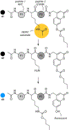
Trans-esterification HDAC probes. This K4(Ac)-CBB probe design contains acetylated peptide that acts as an HDAC substrate and a fluorescent response element appended to its C-terminus. When exposed to an HDAC, hydrolysis results in release of a primary amine, which can undergo an intramolecular trans-esterification with a carbonate ultimately releasing the coumarin 43, which now becomes fluorescent due to increased charge transfer between the free hydroxyl group and the coumarin.
Figure 27.
Figure 27.
Methods to map HDAC drug target complexes using a chemoproteomics competition-binding assay to profile HDAC inhibitor target complexes. This process begins by preparing a probe matrix by derivatizing Sepharose with analogues of nonselective HDAC inhibitors for SAHA in a) and givinostat in b).
Figure 28.
Figure 28.
Illustration of the Paal-Knorr target-guided synthesis strategy. The binding of an aniline (yellow) and 1,4-diketone (blue) within the active site of COX-2 enables an in situ Paal-Knorr reaction that delivers a pyrrole product whose structure can be tailored to be fluorescent by the use of aryl-substituted diketones and aromatic amines, as depicted in Figure 29.
Figure 29.
Figure 29.
Structure of fluorescent pyrroles 46a, 46b, and 46c. Pyrrole 46a was synthesized at the COX-2 active site using aniline 47 and diketone 48. The latter was generated from the Stetter reaction of aldehyde 49 with methylvinyl ketone (50).
Figure 30.
Figure 30.
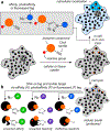
Chemical proteomics. Comparable to conventional mode of action (MOA) workflows, the advance of chemical proteomics has enabled rapid elucidation of small molecule targets in cells and organisms. Schematically, this process begins with the addition of a Click handle (illustrated by alkyne) to the compound (orange sphere) this is followed by appendage of a covalently reactive group (when possible). a) The probe is then presented to the cell, tissue or organism and in cell Click chemistry can be used to track the cellular up take and subcellular localization. b) The molecular targets are then identified by appending either an affinity (A, green), photoaffinity (P, purple) or fluorescence (F) tag and isolating the proteins chromatographically using affinity resins or 2D SDS-PAGE gels.
Figure 31.
Figure 31.
Importance of controls in centralized workflows. The development or uniform linker and controls are critical to studies that explore the cellular and molecular mode and mechanisms of a compound’s activity. a) In this study, a fluorescent coumarin analogue 51 was prepared as a replacement of the alkene terminus of 33-OMe-phorboxazole B (52). b) Both cellular localization and molecular targeting was identified by comparing the activity of 53 to a fluorescent linker control 51. Here, different control analogues were included to expand the SAR of the controls to ensure that the fluorescent tag and its conjugation within51 do not present off-target activity.
Figure 32.
Figure 32.
Bidirectional affinity natural product Isolation. A five-step protocol has been reported that enables one to agnostically-discover natural products and their associated protein targets (step 1). The procedure begins by preparation of proteomic resins by lysing targeted cells and covalently-attaching the resulting proteins to reactive resins such as Affi-Gel 10. Step 2: The resulting proteomic resins are then used to as affinity matrixes to isolate natural products from crude or fractionated extracts. Step 3: The proteome bound natural products where then tagged with an immunoaffinity fluorescent (IAF) tag (54) and purified using resins bearing an antibody against the IAF tag (55). Step 4: The resulting probes were then used for uptake and subcellular localization studies and (step 5) target immunoprecipitation using resin bound antibodies against the IAF tag.
Figure 33.
Figure 33.
Inverse drug discovery strategy. Recently a team at Scripps Research described an approach to drug discovery that united covalently reactive compounds, click tags for detection, proteomic analyses and structural biology to identify binding pockets for therapeutic discovery. In this example a reactive fluorosulfate is used to append a clickable tag for SDS PAGE proteomic analyses. One protein GSTO01 was identified and the structure of its covalent adduct was determined by X-ray crystallography. A clicked fluorescent Click tag enabled the detection and isolation of the targeted protein. While the structure of its tag was not critical to the effort, the use of this tag illustrates an important application for probe development.
Figure 34.
Figure 34.
Nanoscale synthesis and affinity ranking (nanoSAR). A schematic representation of the reaction and chemical space prepared and biologically assayed directly to identify potent inhibitors and reaction conditions, simultaneously. Nu represents building blocks that were coupled to 56 by a Suzuki and C–N couplings. A total of 170 reactions were conducted to prepare and screen a library of 20 CHK1 inhibitors. A total of 8.8 mg of 56 was used for this study. Reaction conditions included a catalyst (tBuBrettPhos Pd G3), base (7-methyl-1,5,7-triazabicyclo[4.4.0]dec-5-ene or MTBD) in 1-methyl-2-pyrrolidinone (NMP, solvent). Each reaction was run at nanomol scales and leads were rescaled at the 50 μmol scale. While the structure of its fluorophore was not critical to the effort, this study illustrates an excellent application for future probe designs.
Figure 35.
Figure 35.
Target protein-guided hit prioritization and probe identification. Two different resins, a) a scanning confocal microscopy (CONA) resin and b) a functional chromatographic (FC) resin, are applied to screen extracts A–B for protein binders and isolate them. The CONA resin us used to prioritize the extracts at high-throughput and the second or FC resin is used to isolate the target protein binding compounds. Using 1.7 mm capillary NMR the team was able to conduct this dual-resin affinity screen using to discover probes as low as 5 μg of material.
Figure 36.
Figure 36.
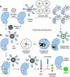
Isotope-coded fluorogenic crosslinking, a schematic representation of the fPAL strategy. A two-step process involves carbene activation and photochemical target capture results in a fluorescent adduct 61 from 59. Intramolecular attack by a proximal phenol within 60 results in the formation of a fluorescent coumarin motif, which is covalently attached to an amino acid residue within the binding pocket of the targeted protein.
Figure 37.
Figure 37.
Acyl fluorophore transfer concept. a) A schematic representation of the strategy, wherein a fluorophore-transfer cassette 62. provides both a fluorophore and ligand (a natural product). Binding of a protein target results in a reaction between a nucleophilic residues in the active site of the bound complex 63. This results in the loss of the natural product (a leaving group) and fluorescent tagging of the target protein within its binding pocket. b) Structures of marinopyrrole A (65) and the fluorophore-transfer cassette 66 prepared in this study.
Figure 38.
Figure 38.
Tosyl chemistry for labeling endogenous proteins in living native cells. a) Schematic illustration of the strategy. The method allows for the labeling of a target protein with a fluorescent (or affinity) tag within its binding pocket. b) Probe 70 contained a central fluorophore-cassette that was linked to a fluorophore and a ligand. In this example the ligand was designed to target carbonic anhydrase.
Figure 39.
Figure 39.
Conversion or aromatic diamine to triazole through a reaction with nitric oxide in the presence of oxygen. Diamines 71 and 72 are not fluorescent because of the photoinduced electron transfer quenching from the electron-rich amino groups. The transformation of the diamino moiety to the triazole reduces the electron-donating ability of the nitrogen atoms reducing fluorescence quenching of the fluorophore moiety and resulting in a turn-on emission. The technology has been applied to cyanine 71 and BODIPY 72 scaffolds as well as diaminorhodamine 75 and diaminofluorescein 76. Circles for 71 and 73 provide IR wavelengths numerically in nm.
Figure 40.
Figure 40.
Proposed mechanism involved in the detection of protease activity based on the covalent-assembly approach. Leucine aminopeptidase 77 (LAP) hydrolyses the amide bond and releases aniline. Cyclization followed by the hydroxyl departure gives fluorescent pyronin 81.
Figure 41.
Figure 41.
Hg2+-promoted cyclization of thiosemicarbazides to 1,3,4-oxadiazoles leads to irreversible conversion of spirolactam 82 to fluorescent ring-opened rhodamine derivative 83. When mercury ions are added to a PBS solution of 82, the latter instantaneously turns from colorless to pink and becomes strongly fluorescent.
Figure 42.
Figure 42.
Near IR fluorophores. a) Structure of IR-780, a NIR fluorophore with significant accumulation in mitochondria of tumor cells. b) Structure of IR-780/nitrogen mustard theranostic agent showing excellent accumulation in tumor xenografts in vivo. Circles for 84 and 85 provide IR wavelengths numerically in nm.

Similar articles

Cited by

References

    1. Monardes N in Historia medicinal de las cosas que se traen de nuestras Indias Occidentales, Seville, 1565.
    1. Sahagún B in 1499–1590, Florentine Codex: General History of the Things of New Spain, 2nd ed., Revised, Santa Fe, N. M.: The School Of American Research; Salt Lake City, Utah, University Of Utah, 1970.
    1. Acuña AU, Amat-Guerri F, Morcillo P, Liras M, Rodriguez B, Org. Lett 2009, 11, 3020–3023; - PubMed
    2. Tomalia DA, Klajnert-Maculewicz B, Johnson KA-M, Brinkman HF, Janaszewska A, Hedstrand DM, Prog. Polym. Sci 2019, 90, 35–117.
    1. Acuña AU, Amat-Guerri F, in Fluorescence of Supermolecules, Polymers, and Nanosystems, Springer, 2007, pp. 3–20.
    1. Specht EA, Braselmann E, Palmer AE, Annu. Rev. Physiol 2017, 79, 93–117. - PubMed

Publication types