. 2021 Dec;59(1):662-671.
doi: 10.1080/13880209.2021.1928241.

Angelica gigas root ameliorates ischaemic stroke-induced brain injury in mice by activating the PI3K/AKT/mTOR and MAPK pathways

Affiliations

Angelica gigas root ameliorates ischaemic stroke-induced brain injury in mice by activating the PI3K/AKT/mTOR and MAPK pathways

Se-Eun Lee et al. Pharm Biol. 2021 Dec.

Abstract

Context: Traditionally, the root of Angelica gigas Nakai (Umbelliferae), has long been used to treat ischaemic diseases and is considered safe in humans.

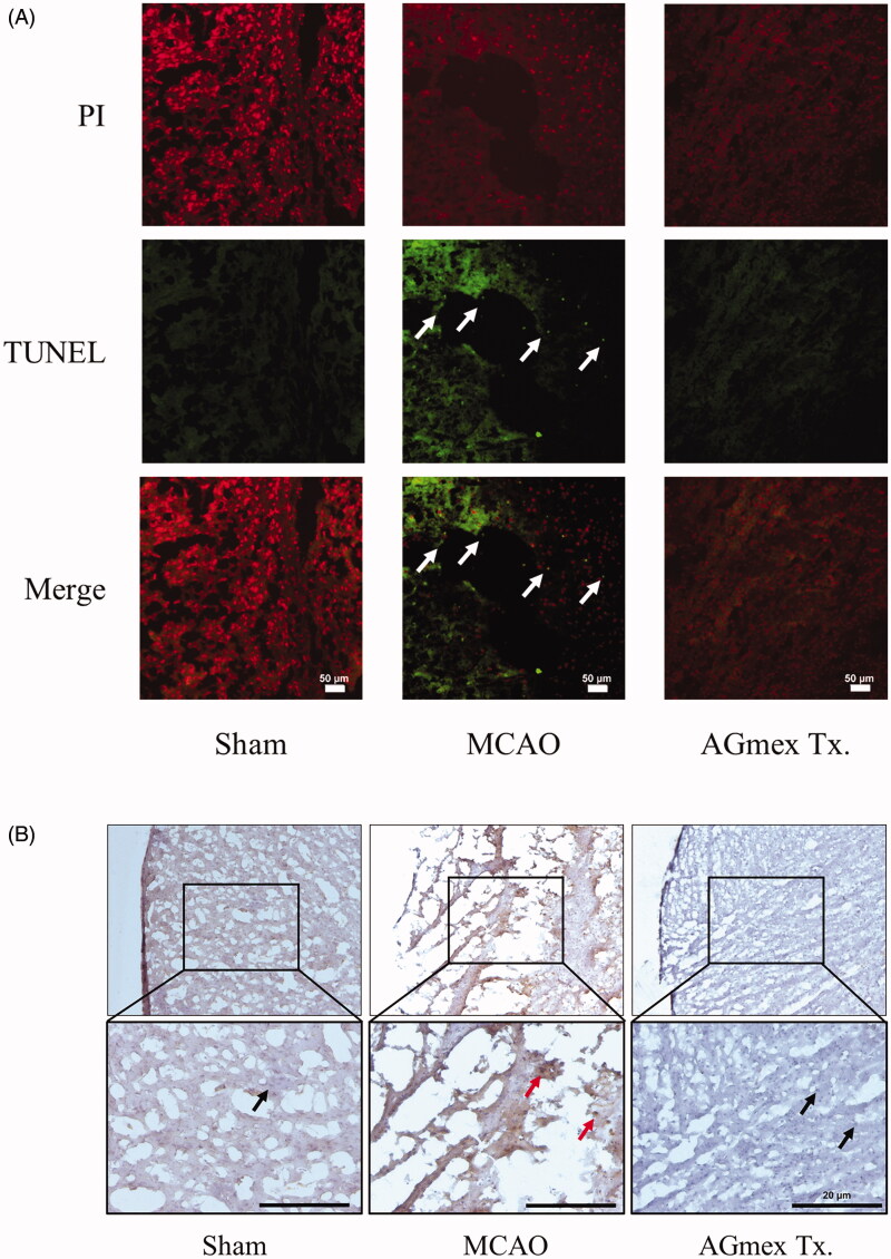
Objective: To investigate the neuroprotective effects of a methanol extract of A. gigas root (AGmex) on the middle cerebral artery occlusion (MCAO)-induced brain injury in mice, and the underlying mechanisms.

Materials and methods: Two hours of transient MCAO (tMCAO) was induced in C57BL/6 mice (MCAO control group and AGmex groups), AGmex was administered to the AGmex group at 300-3,000 mg/kg bw at 1, 1, and 24 h before tMCAO or at 1000 mg/kg bw at 1 h before and after tMCAO. Infarction volumes, tissue staining, and western blotting were used to investigate the mechanism underlying the neuroprotective effects of AGmex.

Results: The median effective dose (ED50) could not be measured because the AGmex treatment did not reduce the infarction volume caused by 2 h of tMCAO to within 50%; however, pre-treatment with AGmex twice at 1,000 mg/kg bw before tMCAO significantly reduced the infarction volumes. The proteins related to cell growth, differentiation, and death were upregulated by this treatment, and the major recovery mechanisms appeared to involve the attenuation of the mitochondrial function of Bcl-2/Bax and activation of the PI3K/AKT/mTOR and MAPK signalling pathways in ischaemic neurons.

Conclusions: This study provides evidence supporting the use of A. gigas root against ischaemic stroke and suggests a novel developmental starting point for the treatment of ischaemic stroke.

Keywords: Middle cerebral artery occlusion; brain edoema; infarction; neuroprotection.


Conflict of interest statement

No potential conflict of interest was reported by the author(s).

Figures

Figure 1.
Figure 1.
High-performance liquid chromatography (HPLC) results for the methanolic extract of Angelica gigas root (AGmex) and the nodakenin and decursin standards. A, HPLC chromatogram of the standards and their chemical structures. B, HPLC chromatogram of AGmex. C, Mobile phase gradient (mobile phase A, acetonitrile; mobile phase B, water). Conditions used: HPLC, Shimadzu system (Shimadzu, Kyoto, Japan); column, YMC-Triart C18; wavelength, 330 nm; column temperature, 35 °C; flow rate, 1 mL/min; injection volume, 10 μL.
Figure 2.
Figure 2.
Experimental schedule of the 2 h transient middle cerebral artery occlusion (tMCAO) model. The mice were acclimated for 1 week in our animal facility before study commencement. Pre-treatment (Pre-Tx.) and post-treatment (Post-Tx.) studies were carried out to determine the timing of AGmex administration. TTC; 2,3,5-triphenyl-tetrazolium chloride; EB; Evans blue.
Figure 3.
Figure 3.
Infarct volumes, edoema areas, and neurologic deficit scores (NDS). (A) Representative photographs of TTC-stained brain coronal slices (1 mm) showing infarct areas 24 h after tMCAO (upper column) and total infarct volumes (lower column). (B) Quantitative analysis of the total edoema area. (C) Neurologic deficit score (NDS). S = single AGmex treatment at 300, 1000 or 3000 mg/kg bw p.o. and T = AGmex treatment for two consecutive days at 300, 1000 or 3000 mg/kg bw/d p.o.; ND, nimodipine (positive control), which was administered once or twice at 60 mg/kg at 1 h or 1 h and 24 h after MCAO, respectively. Results are presented as means ± SDs (n = 3). ##p < 0.01, ###p < 0.001 vs. the sham-operated group; *p < 0.05, **p < 0.01, ***p < 0.001 vs. the MCAO group.
Figure 4.
Figure 4.
Effects of AGmex on blood-brain barrier (BBB) permeability and the localisation of AQP4 (aquaporin 4) protein in MCAO-induced mouse brains. (A) Representative images of EB extravasation in whole brain and coronal sections (upper column), and quantification of EB leakage in the ipsilateral (left) hemispheres and contralateral (right) hemispheres (lower column, n = 3). (B), Immunohistochemical (IHC) staining of AQP4 protein in the cerebral cortex (black arrows indicate negativity and red arrows positivity). Scale bars: 5 µm. Results are presented as means ± SDs (n = 3). ###p < 0.001 vs. the sham-operated group; *p < 0.05 and ***p < 0.001 vs. the MCAO group.
Figure 5.
Figure 5.
Neuroprotective effects of double pre-treatment (at 1 h and 24 h pre-MCAO) with AGmex at 1000 mg/kg on MCAO-induced cell death. (A) Representative photomicrographs of Nissl-stained neurons in the subcortical area (upper images) ischaemic ipsilateral hemisphere and results of quantitative analysis showing changes in neuron numbers following tMCAO induction and pre-treatment with AGmex (lower graph). (B) Representative photomicrographs of haematoxylin and eosin (H&E)-stained neurons (upper images) and results of quantitative analysis showing changes in neuron numbers (lower graph). Black arrows indicate intact neurons with normal morphology, and red arrows indicate neurons showing apoptotic changes and aberrant morphologies. Scale bars: 20 µm. #p < 0.05, ##p < 0.01 vs. sham controls; *p < 0.05, **p < 0.01 vs. the MCAO group.
Figure 6.
Figure 6.
Effects of double pre-treatment (at 1 h and 24 h pre-MCAO) with AGmex at 1000 mg/kg on MCAO-induced cell death. (A) Representative images of TUNEL and PI double-stained cerebral cortex regions showing MCAO-induced apoptosis; white arrows indicate neurons showing apoptotic changes (scale bars: 50 µm). (B) Representative images of IHC staining of MCAO-induced changes in mTOR (mammalian target of rapamycin) protein expression in the cerebral subcortical regions of tMCAO-induced mice; red arrows indicate mTOR positivity, and black arrows indicate mTOR negativity (scale bars: 20 µm).
Figure 7.
Figure 7.
Effects of double pre-treatment (at 1 h and 24 h pre-MCAO) with AGmex at 1000 mg/kg on the proteins expressed in MCAO-induced mouse brains. (A) Representative western blot image of the expression of Bcl-2, Bax, p-PI3K, PI3K, p-AKT, AKT, p-ERK, ERK, p-JNK, JNK, p-p38, p38, SIRT1, MnSOD, and β-actin in MCAO-treated mouse brain. B-I, ratios of the expression levels of signalling proteins: (B) Bcl-2/Bax; C, p-PI3K/PI3K; D, p-AKT/AKT; E, p-ERK/ERK; F, p-JNK/JNK; G, p-p38/p38; H, SIRT1/β-actin; and I, MnSOD/β-actin. Results are presented as means ± SDs (n = 3). #p < 0.05, ###p < 0.001 vs. sham-operated controls; *p < 0.05, **p < 0.01 vs. the MCAO group.
Figure 8.
Figure 8.
Schematic view of the suggested cerebral damage pathways and the neuroprotective mechanism of AGmex. AGmex ameliorated mitochondrial function and MAPK signalling pathways in the neurons exposed to ischaemia, and the neuroprotective effects of AGmex were found to mainly involve anti-edoema and anti-apoptotic pathways.

Similar articles

Cited by

References

    1. Ambe K, Watanabe H, Takahashi S, Nakagawa T.. 2014. Immunohistochemical localization of Nox1, Nox4 and Mn-SOD in mouse femur during endochondral ossification. Tissue Cell. 46:433–438. - PubMed
    1. Badaut J, Fukuda AM, Jullienne A, Petry KG.. 2014. Aquaporin and brain diseases. Biochim Biophys Acta. 1840:1554–1565. - PMC - PubMed
    1. Cao G, Pei W, Lan J, Stetler RA, Luo Y, Nagayama T, Graham SH, Yin XM, Simon RP, Chen J.. 2001. Caspase-activated DNase/DNA fragmentation factor 40 mediates apoptotic DNA fragmentation in transient cerebral ischemia and in neuronal cultures. Neuroscience. 21:4678–4690. - PMC - PubMed
    1. Cho JH, Kwon JE, Cho Y, Kim I, Kang SC.. 2015. Anti-inflammatory effect of Angelica gigas via heme oxygenase (HO)-1 expression. Nutrients. 7:4862–4874. - PMC - PubMed
    1. Choi HS, Cho SG, Kim MK, Kim MS, Moon SH, Kim IH, Ko SG.. 2016. Decursin in Angelica gigas Nakai (AGN) enhances doxorubicin chemosensitivity in NCI/ADR-RES ovarian cancer cells via inhibition of p-glycoprotein expression. Phytother Res. 30:2020–2026. - PubMed

MeSH terms