

Clinical Features and Diagnosis of Cardiac Sarcoidosis
- PMID: 34062709
- PMCID: PMC8124502
- DOI: 10.3390/jcm10091941
Clinical Features and Diagnosis of Cardiac Sarcoidosis
Abstract
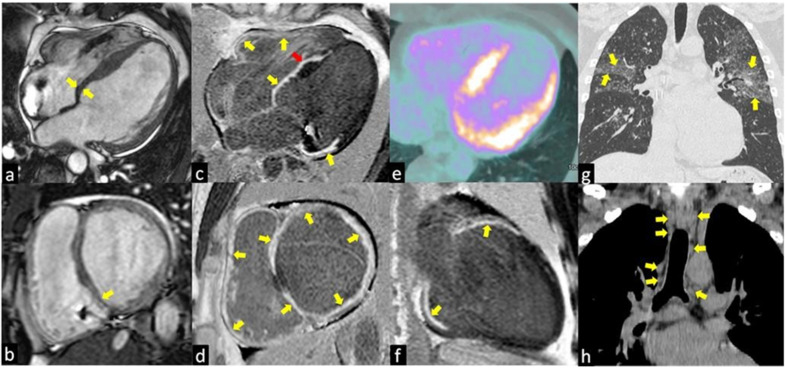
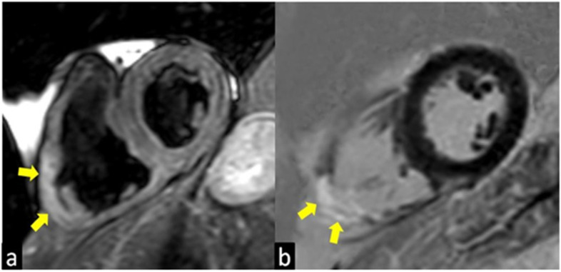
Cardiac sarcoidosis (CS) is an unusual, but potentially harmful, manifestation of systemic sarcoidosis (SA), a chronic disease characterized by organ involvement from noncaseating and nonnecrotizing granulomas. Lungs and intrathoracic lymph nodes are usually the sites that are most frequently affected, but no organ is spared and CS can affect a variable portion of SA patients, up to 25% from post-mortem studies. The cardiovascular involvement is usually associated with a bad prognosis and is responsible for the major cause of death and complications, particularly in African American patients. Furthermore, the diagnosis is often complicated by the occurrence of non-specific clinical manifestations, which can mimic the effect of more common heart disorders, and imaging and biopsies are the most valid approach to avoid misdiagnosis. This narrative review summarizes the main clinical features of CS and imaging findings, particularly of CMR and 18-Fluorodeoxyglucose Positron Emission Tomography (18F-FDG PET) that can give the best cost/benefit ratio in terms of the diagnostic approach. Imaging can be very useful in replacing the endomyocardial biopsy in selected cases, to avoid unnecessary, and potentially dangerous, invasive maneuvers.
Keywords: 18F-FDG PET; biopsy; cardiac sarcoidosis; imaging; magnetic resonance imaging.
Conflict of interest statement
The author declares no conflict of interest.
Figures
Similar articles
-
F-18-fluorodeoxyglucose positron emission tomography-guided sampling of mediastinal lymph nodes in the diagnosis of cardiac sarcoidosis.Am J Cardiol. 2015 Nov 15;116(10):1581-5. doi: 10.1016/j.amjcard.2015.08.025. Epub 2015 Sep 3. Am J Cardiol. 2015. PMID: 26411357
-
Comparison of (18)F-fluorodeoxyglucose positron emission tomography (FDG PET) and cardiac magnetic resonance (CMR) in corticosteroid-naive patients with conduction system disease due to cardiac sarcoidosis.Eur J Nucl Med Mol Imaging. 2016 Feb;43(2):259-269. doi: 10.1007/s00259-015-3181-8. Epub 2015 Sep 11. Eur J Nucl Med Mol Imaging. 2016. PMID: 26359191
-
PET/CT in the Diagnosis and Workup of Sarcoidosis: Focus on Atypical Manifestations.Radiographics. 2018 Sep-Oct;38(5):1536-1549. doi: 10.1148/rg.2018180053. Epub 2018 Aug 17. Radiographics. 2018. PMID: 30118393 Review.
-
Characteristics and survival of patients diagnosed with cardiac sarcoidosis: A case series.Front Med (Lausanne). 2022 Dec 13;9:1051412. doi: 10.3389/fmed.2022.1051412. eCollection 2022. Front Med (Lausanne). 2022. PMID: 36582282 Free PMC article.
-
[Cardiac sarcoidosis - clinical manifestation and diagnosis].Pol Merkur Lekarski. 2016 Aug;41(242):101-6. Pol Merkur Lekarski. 2016. PMID: 27591449 Review. Polish.
Cited by
-
Clinical Features, Histopathology and Differential Diagnosis of Sarcoidosis.Cells. 2021 Dec 26;11(1):59. doi: 10.3390/cells11010059. Cells. 2021. PMID: 35011621 Free PMC article. Review.
-
Exploring the latest advances in 18F-FDG PET/CT and cardiac magnetic resonance for imaging for cardiac sarcoidosis diagnosis.Am J Nucl Med Mol Imaging. 2024 Apr 25;14(2):149-156. doi: 10.62347/GIKK5707. eCollection 2024. Am J Nucl Med Mol Imaging. 2024. PMID: 38737647 Free PMC article. Review.
-
Comorbidities of sarcoidosis.Ann Med. 2022 Dec;54(1):1014-1035. doi: 10.1080/07853890.2022.2063375. Ann Med. 2022. PMID: 35441568 Free PMC article. Review.
-
Sarcoidosis-associated pulmonary hypertension due to pulmonary arteries stenosis - a case report.BMC Pulm Med. 2024 Jul 16;24(1):346. doi: 10.1186/s12890-024-03152-0. BMC Pulm Med. 2024. PMID: 39014431 Free PMC article.
-
Neuropsychiatric manifestations of sarcoidosis.Ann Med. 2025 Dec;57(1):2445191. doi: 10.1080/07853890.2024.2445191. Epub 2024 Dec 26. Ann Med. 2025. PMID: 39723989 Free PMC article. Review.
References
-
- Tana C., Tchernev G., Chokoeva A.A., Wollina U., Lotti T., Fioranelli M., Roccia M.G., Maximov G.K., Silingardi M. Pulmonary and abdominal sarcoidosis, the great imitators on imaging? J. Biol. Regul. Homeost. Agents. 2016;30:45–48. - PubMed
Publication types
LinkOut - more resources
Full Text Sources
Research Materials
