

Robust and Accurate Mandible Segmentation on Dental CBCT Scans Affected by Metal Artifacts Using a Prior Shape Model
- PMID: 34062762
- PMCID: PMC8147374
- DOI: 10.3390/jpm11050364
Robust and Accurate Mandible Segmentation on Dental CBCT Scans Affected by Metal Artifacts Using a Prior Shape Model
Abstract
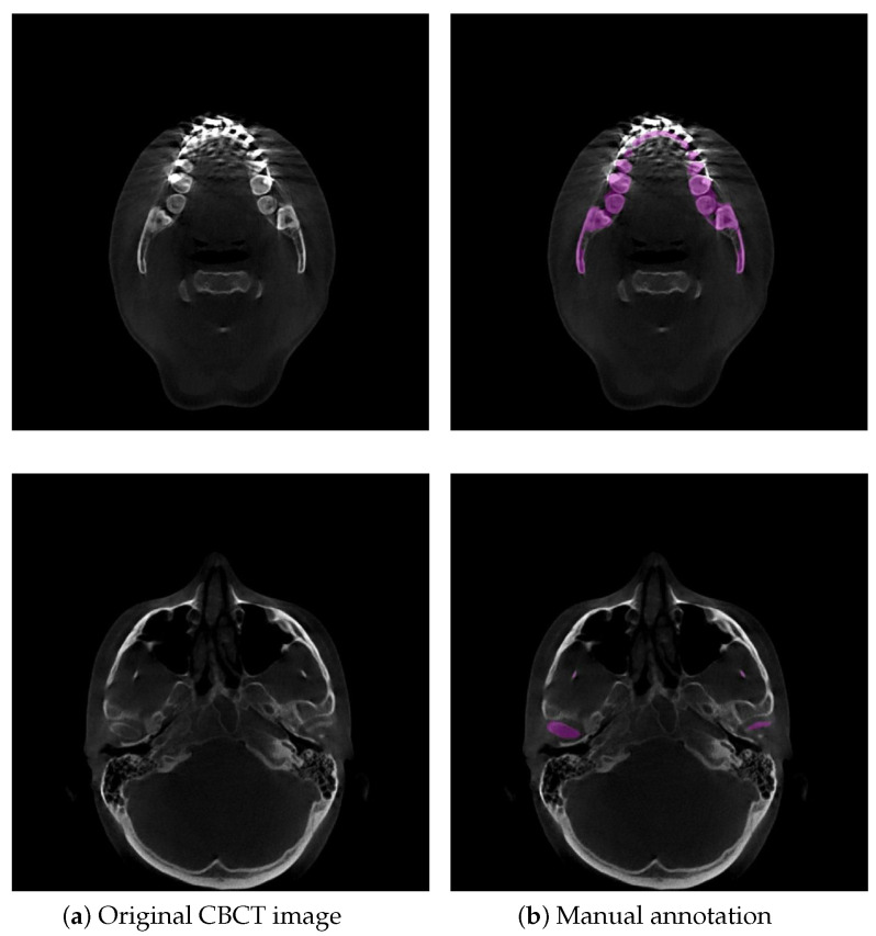
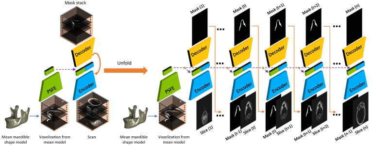
Accurate mandible segmentation is significant in the field of maxillofacial surgery to guide clinical diagnosis and treatment and develop appropriate surgical plans. In particular, cone-beam computed tomography (CBCT) images with metal parts, such as those used in oral and maxillofacial surgery (OMFS), often have susceptibilities when metal artifacts are present such as weak and blurred boundaries caused by a high-attenuation material and a low radiation dose in image acquisition. To overcome this problem, this paper proposes a novel deep learning-based approach (SASeg) for automated mandible segmentation that perceives overall mandible anatomical knowledge. SASeg utilizes a prior shape feature extractor (PSFE) module based on a mean mandible shape, and recurrent connections maintain the continuity structure of the mandible. The effectiveness of the proposed network is substantiated on a dental CBCT dataset from orthodontic treatment containing 59 patients. The experiments show that the proposed SASeg can be easily used to improve the prediction accuracy in a dental CBCT dataset corrupted by metal artifacts. In addition, the experimental results on the PDDCA dataset demonstrate that, compared with the state-of-the-art mandible segmentation models, our proposed SASeg can achieve better segmentation performance.
Keywords: 3D virtual surgical planning (3D VSP); accurate mandible segmentation; convolutional neural network; oral and maxillofacial surgery.
Conflict of interest statement
The authors declare no conflict of interest.
Figures


Similar articles
-
Mandible Segmentation of Dental CBCT Scans Affected by Metal Artifacts Using Coarse-to-Fine Learning Model.J Pers Med. 2021 Jun 16;11(6):560. doi: 10.3390/jpm11060560. J Pers Med. 2021. PMID: 34208429 Free PMC article.
-
Recurrent Convolutional Neural Networks for 3D Mandible Segmentation in Computed Tomography.J Pers Med. 2021 May 31;11(6):492. doi: 10.3390/jpm11060492. J Pers Med. 2021. PMID: 34072714 Free PMC article.
-
Segmentation of dental cone-beam CT scans affected by metal artifacts using a mixed-scale dense convolutional neural network.Med Phys. 2019 Nov;46(11):5027-5035. doi: 10.1002/mp.13793. Epub 2019 Sep 13. Med Phys. 2019. PMID: 31463937 Free PMC article.
-
Automatic Segmentation of Mandible from Conventional Methods to Deep Learning-A Review.J Pers Med. 2021 Jul 1;11(7):629. doi: 10.3390/jpm11070629. J Pers Med. 2021. PMID: 34357096 Free PMC article. Review.
-
Cone beam computed tomography in implant dentistry: recommendations for clinical use.BMC Oral Health. 2018 May 15;18(1):88. doi: 10.1186/s12903-018-0523-5. BMC Oral Health. 2018. PMID: 29764458 Free PMC article. Review.
Cited by
-
Morphological Variation of the Mandible in the Orthognathic Population-A Morphological Study Using Statistical Shape Modelling.J Pers Med. 2023 May 19;13(5):854. doi: 10.3390/jpm13050854. J Pers Med. 2023. PMID: 37241024 Free PMC article.
-
Use of Advanced Artificial Intelligence in Forensic Medicine, Forensic Anthropology and Clinical Anatomy.Healthcare (Basel). 2021 Nov 12;9(11):1545. doi: 10.3390/healthcare9111545. Healthcare (Basel). 2021. PMID: 34828590 Free PMC article.
-
Mandible Segmentation of Dental CBCT Scans Affected by Metal Artifacts Using Coarse-to-Fine Learning Model.J Pers Med. 2021 Jun 16;11(6):560. doi: 10.3390/jpm11060560. J Pers Med. 2021. PMID: 34208429 Free PMC article.
-
Analysis of Deep Learning Techniques for Dental Informatics: A Systematic Literature Review.Healthcare (Basel). 2022 Sep 28;10(10):1892. doi: 10.3390/healthcare10101892. Healthcare (Basel). 2022. PMID: 36292339 Free PMC article. Review.
-
Accuracy of artificial intelligence-based segmentation in maxillofacial structures: a systematic review.BMC Oral Health. 2025 Mar 7;25(1):350. doi: 10.1186/s12903-025-05730-y. BMC Oral Health. 2025. PMID: 40055718 Free PMC article.
References
-
- Kraeima J. Ph.D. Thesis. University of Groningen; Groningen, The Netherlands: 2019. Three Dimensional Virtual Surgical Planning for Patient Specific Osteosynthesis and Devices in Oral In addition, Maxillofacial Surgery. A New Era.
-
- Vaitiekūnas M., Jegelevičius D., Sakalauskas A., Grybauskas S. Automatic Method for Bone Segmentation in Cone Beam Computed Tomography Data Set. Appl. Sci. 2020;10:236. doi: 10.3390/app10010236. - DOI
LinkOut - more resources
Full Text Sources
