Review
. 2021 May 1;13(9):2184.
doi: 10.3390/cancers13092184.

Natural Killer-Dendritic Cell Interactions in Liver Cancer: Implications for Immunotherapy

Affiliations
Review

Natural Killer-Dendritic Cell Interactions in Liver Cancer: Implications for Immunotherapy

Valentina Cazzetta et al. Cancers (Basel). .

Abstract

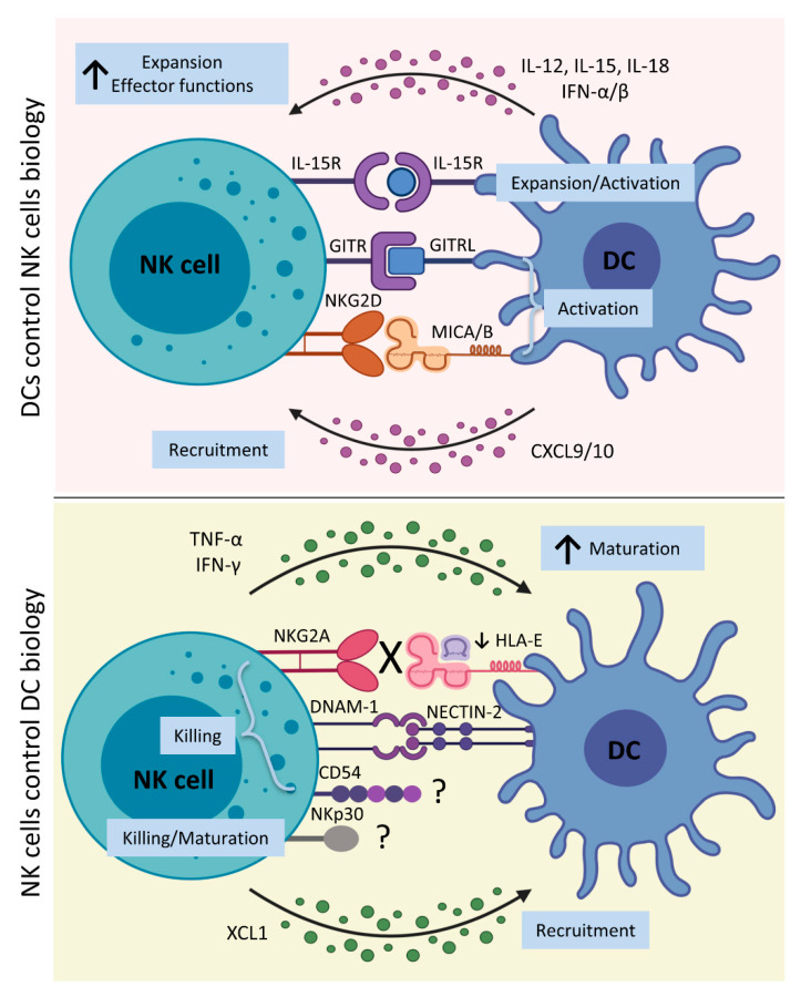
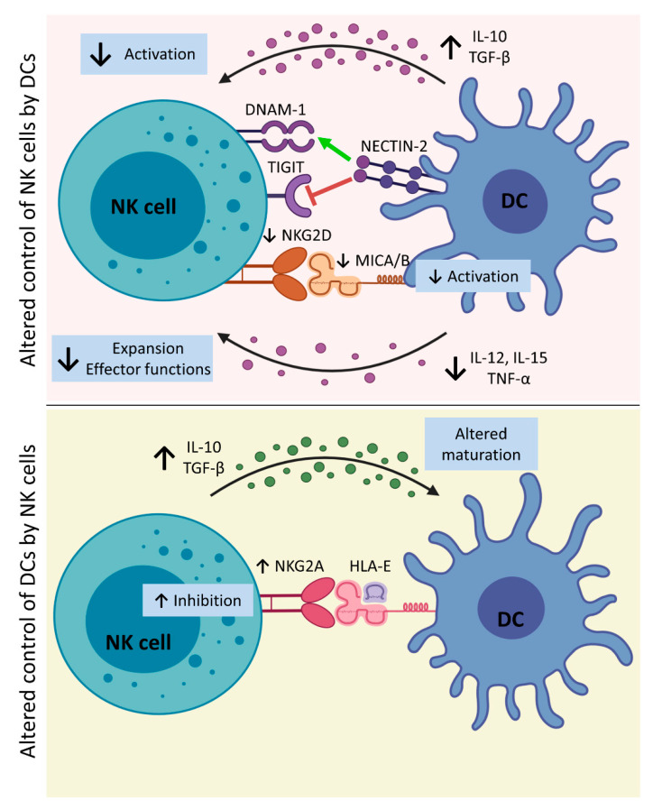
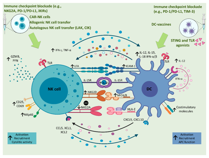
Natural killer (NK) and dendritic cells (DCs) are innate immune cells that play a crucial role in anti-tumor immunity. NK cells kill tumor cells through direct cytotoxicity and cytokine secretion. DCs are needed for the activation of adaptive immune responses against tumor cells. Both NK cells and DCs are subdivided in several subsets endowed with specialized effector functions. Crosstalk between NK cells and DCs leads to the reciprocal control of their activation and polarization of immune responses. In this review, we describe the role of NK cells and DCs in liver cancer, focusing on the mechanisms involved in their reciprocal control and activation. In this context, intrahepatic NK cells and DCs present unique immunological features, due to the constant exposure to non-self-circulating antigens. These interactions might play a fundamental role in the pathology of primary liver cancer, namely hepatocellular carcinoma (HCC) and intrahepatic cholangiocarcinoma (ICC). Additionally, the implications of these immune changes are relevant from the perspective of improving the cancer immunotherapy strategies in HCC and ICC patients.

Keywords: DCs–NK cell interaction; cancer therapy; innate immunity; liver cancer.


Conflict of interest statement

The authors declare no conflict of interest.

Figures

Figure 1
Figure 1
DC–NK cell interplay. Upper panel: After exposure to several stimuli recognized by a wide variety of pattern recognition receptors expressed by DCs, they release different cytokines, like IL-12, IL-15, IL-18, and IFN-γ. IL-15 trans-presentation by DCs is required for NK cell priming, and IL-15 cis-presentation by NK cells contributes to NK cell activation through the production of IFN-β by DCs. IL-12 and IL-18 stimulate NK cells to produce IFN-γ and enhance NK cell cytotoxicity. Moreover, direct NK–DC contact is required for optimal NK cell activation by DCs. The interaction of NKG2D with MICA/B expressed by DCs upon IFN-α simulation, and the interaction of GITR with its ligand on DCs also lead to NK cell activation. Moreover, DCs release chemokines, such as CXCL9/10, which further recruit NK cells at the site of inflammation. Lower panel: Activated NK cells produce IFN-γ and TNF-α, which promote maturation of DCs that are able to drive Th1- and cytotoxic T cell development. Additionally, the NKp30 engagement is involved in this process. To increase DC recruitment at the site of inflammation, NK cells release XCL1, which binds to XCR1 expressed by DCs. To limit the supply of DCs at sites of inflammation and control excessive adaptive immune response, NK cells that express the inhibitory receptor NKG2A and lack activating KIRs can kill autologous iDCs that display a reduction in HLA-E expression. In particular, the direct NK–DC interaction, through NKp30, DNAM-1, and CD54, on NK cell surface, and their ligands on DC surface, induces the killing of iDCs. Notably, NKp30 and CD54 ligands on DCs surface are still unknown, as indicated in the figure by a question mark. [CXCL10, Chemokine (C-X-C motif) ligand 10; CXCL9, Chemokine (C-X-C motif) ligand 9; DCs, dendritic cells; DNAM-1, DNAX Accessory Molecule-1 or CD226; GITR, glucocorticoid-induced tumor necrosis factor receptor; GITRL, glucocorticoid-induced tumor necrosis factor receptor ligand; HLA-E, Major Histocompatibility Complex, Class I, E; iDCs, immature dendritic cells; IFN-α, interferon α; IFN-β, interferon β; IFN-γ, interferon γ; IL-12, interleukin 12; IL-15, interleukin 15; IL-15R, interleukin 15 receptor; IL-18, interleukin 18; KIRs, Killer-cell immunoglobulin-like receptors; MICA/B, MHC class-I-related protein A/B; NECTIN-2, Nectin Cell Adhesion Molecule 2 or CD112; NK cells, natural killer cells; TNF-α, tumor necrosis factor α; XCL1, Chemokine (C motif) ligand 1; and XCR1, X-C Motif Chemokine Receptor 1]. Created with BioRender.com (accessed on 16 March 2021).
Figure 2
Figure 2
Altered DC–NK cell interplay in liver cancer. Upper panel: In the TME, DCs increase the secretion of IL-10 and TGF-β, which impair NK cell activation by the reduction of NKG2D expression on NK cells. Therefore, altered NKG2D–MICA/B interaction results in lower NK cell and DC activation. Moreover, the exposure to high levels of IL-10 and TGF-β induces a lower release of IL-12, IL-15, and TNF-α by DCs. In particular, impaired production of IL-15, causes the aberrant expression of MICA/B on DCs, and decreased expression of IL-12 by DCs induced by tumor AFP further harm NK cell activation. NK cell interaction by NECTIN-2 exposed on DCs can either block or increase NK cell activity through TIGIT or DNAM-1 interaction, respectively. Lower panel: Tumor-associated NK cells increase expressions of IL-10 and TGF-β that impair DC maturation, leading to the killing of iDCs by NK cells. Moreover, increased expressions of NKG2A on NK cells and HLA-E on DCs inhibit NK cell activity. [AFP, α-fetoprotein; DCs, dendritic cells; DNAM-1, DNAX Accessory Molecule-1 or CD226; HLA-E, Major Histocompatibility Complex, Class I, E; iDCs, immature dendritic cells; IL-10, interleukin 10; IL-12, interleukin 12; IL-15, interleukin 15; IL-18, interleukin 18; MICA/B, MHC class-I-related protein A/B; NK cells, natural killer cells; NECTIN-2, Nectin Cell Adhesion Molecule 2 or CD112; TGF-β, tumor growth factor β; TIGIT, T cell immunoreceptor with Ig and ITIM domains; TME, tumor microenvironment; and TNF-α, tumor necrosis factor α]. Created with BioRender.com (accessed on 16 March 2021).
Figure 3
Figure 3
Implications of NK–DC crosstalk in liver cancer immunotherapy. Several immunotherapeutic approaches based on the administration of CAR-NK cells, allogenic or autologous NK cells or DC-vaccines are used in cancer patients to activate innate immune responses against tumor cells. The NK cell activation could also be reached through the administration of TLR agonists, like IQ and GDQ, which are, respectively, TLR7 and TLR7/8 agonists. NK cell activation on one hand leads to DC maturation, and on the other hand it is enhanced by the interaction with DCs, which in turn are activated by TLR agonists or STING. This bilateral activation involves the upregulation of CD69, CD25, NKp46, NKG2D, and the release of lytic enzymes (e.g., GZMB and PFN) by NK cells. On the other hand, activated DCs undergo upregulation of costimulatory molecules, NKG2D ligands (e.g., MICA/B) and an increased release of IFN-α, which in a positive loop, also contributes to the production of IL-12 that activates NK cells to produce high levels of IFN-γ and TNF-α. In addition, IL-15 and IL-18 sustain NK cell survival and activation that are also favored by the direct contact between NK cells and DCs through the interaction between NKG2D and its ligands, and LFA and ICAM-1, on NK cell and DC surface, respectively. Moreover, the reciprocal recruitment of both cellular populations is supported by the production of CXCL9 and CXCL10 by DCs and CCL5, XCL1, and XCL2 by NK cells. Other immunotherapeutic approaches, aimed at blocking inhibitory receptors, like the NKG2A, iKIRs, and the PD-1/PD-L1 axis, favor the activation of NK cells, consequently allowing DC maturation and activation, which could be also enhanced by the administration of immune checkpoint inhibitors against PD-1/PD-L1 or TIM-3. [CAR-NK cells, Chimeric Antigen Receptor natural killer cells; CCL5, C-C Motif Chemokine Ligand 5; CXCL10, Chemokine (C-X-C motif) ligand 10; CXCL9, Chemokine (C-X-C motif) ligand 9; DCs, dendritic cells; GDQ, Gardiquimod; GZMB, granzyme B; HLA-E, Major Histocompatibility Complex, Class I, E; IFN-α, interferon α; IFN-β, interferon β; IFN-γ, interferon γ; IL-12, interleukin 12; IL-15, interleukin 15; IL-15R, interleukin 15 receptor; IL-18, interleukin 18; iKIRs, inhibitory Killer-cell immunoglobulin-like receptors; MICA/B, MHC class-I-related protein A/B; IQ, Imiquimod; NK cells, natural killer cells; PD-1, programmed cell death protein 1; PD-L1, programmed death-ligand 1; PFN, perforin; STING, stimulator of interferon genes; TIM-3, T-cell immunoglobulin domain and mucin domain 3; TLR, toll-like receptor; TNF-α, tumor necrosis factor α; XCL1, Chemokine (C motif) ligand 1; XCL2, Chemokine (C motif) ligand 2; and XCR1, X-C Motif Chemokine Receptor 1]. Created with BioRender.com (accessed on 16 March 2021).

Similar articles

Cited by

References

    1. Robinson M.W., Harmon C., O’Farrelly C. Liver Immunology and its role in inflammation and homeostasis. Cell Mol. Immunol. 2016;13:267–276. doi: 10.1038/cmi.2016.3. - DOI - PMC - PubMed
    1. Vivier E., Raulet D.H., Moretta A., Caligiuri M.A., Zitvogel L., Lanier L.L., Yokoyama W.M., Ugolini S. Innate or adaptive immunity? The example of natural killer cells. Science. 2011;331:44–49. doi: 10.1126/science.1198687. - DOI - PMC - PubMed
    1. Chiossone L., Dumas P.Y., Vienne M., Vivier E. Natural killer cells and other innate lymphoid cells in cancer. Nat. Rev. Immunol. 2018;18:671–688. doi: 10.1038/s41577-018-0061-z. - DOI - PubMed
    1. Karre K. Natural killer cell recognition of missing self. Nat. Immunol. 2008;9:477–480. doi: 10.1038/ni0508-477. - DOI - PubMed
    1. Lanier L.L. Up on the tightrope: Natural killer cell activation and inhibition. Nat. Immunol. 2008;9:495–502. doi: 10.1038/ni1581. - DOI - PMC - PubMed