. 2021 May 1;9(5):984.
doi: 10.3390/microorganisms9050984.

Unraveling the Central Role of Sulfur-Oxidizing Acidiphilium multivorum LMS in Industrial Bioprocessing of Gold-Bearing Sulfide Concentrates

Affiliations

Unraveling the Central Role of Sulfur-Oxidizing Acidiphilium multivorum LMS in Industrial Bioprocessing of Gold-Bearing Sulfide Concentrates

Anna Panyushkina et al. Microorganisms. .

Abstract

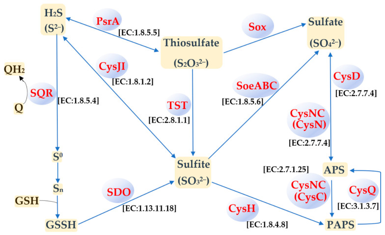
Acidiphilium multivorum LMS is an acidophile isolated from industrial bioreactors during the processing of the gold-bearing pyrite-arsenopyrite concentrate at 38-42 °C. Most strains of this species are obligate organoheterotrophs that do not use ferrous iron or reduced sulfur compounds as energy sources. However, the LMS strain was identified as one of the predominant sulfur oxidizers in acidophilic microbial consortia. In addition to efficient growth under strictly heterotrophic conditions, the LMS strain proved to be an active sulfur oxidizer both in the presence or absence of organic compounds. Interestingly, Ac. multivorum LMS was able to succeed more common sulfur oxidizers in microbial populations, which indicated a previously underestimated role of this bacterium in industrial bioleaching operations. In this study, the first draft genome of the sulfur-oxidizing Ac. multivorum was sequenced and annotated. Based on the functional genome characterization, sulfur metabolism pathways were reconstructed. The LMS strain possessed a complicated multi-enzyme system to oxidize elemental sulfur, thiosulfate, sulfide, and sulfite to sulfate as the final product. Altogether, the phenotypic description and genome analysis unraveled a crucial role of Ac. multivorum in some biomining processes and revealed unique strain-specific characteristics, including the ars genes conferring arsenic resistance, which are similar to those of phylogenetically distinct microorganisms.

Keywords: Acidiphilium multivorum; acidophilic microbial communities; arsenic resistance; biooxidation; gold-bearing sulfide concentrates; sulfur metabolism.


Conflict of interest statement

The authors declare no conflict of interest.

Figures

Figure 1
Figure 1
Dendrogram of Acidiphilium strains based on the 16S rRNA gene sequences, showing the phylogenetic position of Ac. multivorum LMS. The phylogenetic tree was constructed with MEGA X [25]. The evolutionary history was inferred using the Neighbor-Joining method [26]. The percentage of replicate trees in which the associated taxa clustered together in the bootstrap test (1000 replicates) is shown next to the branches [27]. The bootstrap values of ≥80 are shown. The tree is drawn to scale, with branch lengths in the same units as those of the evolutionary distances used to infer the phylogenetic tree. The evolutionary distances were computed using the Maximum Composite Likelihood method [28] and are in the units of the number of base substitutions per site. This analysis involved 11 nucleotide sequences. There were a total of 1511 positions in the final dataset. Kozakia baliensis DSM 14400T was used as an outgroup.
Figure 2
Figure 2
Growth of Ac. multivorum LMS and pH decrease under mixotrophic conditions in the medium supplemented with 0.1% yeast extract and 1% S0.
Figure 3
Figure 3
Micrographs of Ac. multivorum LMS cells grown under mixotrophic conditions in the medium containing 1% S0 and 0.1% yeast extract after isolation from the bioreactor (a) and after 5–7 culture transfers in the same medium (bd). Phase-contrast microscopy. Arrows indicate cell aggregates. S, sulfur. Scale bar, 10 µm.
Figure 4
Figure 4
Alignment of reordered contigs of the Ac. multivorum LMS draft genome and the related reference genome of the type strain Ac. multivorum AIU301T, using the ProgressiveMauve software and Mauve Contig Mover tool [32]. The scale shows sequence coordinates. Colored blocks in the first genome are connected by lines to similarly colored blocks in the second genome. These lines indicate homologous regions in the genomes. Areas that are completely white and not aligned contain sequence elements specific to the genomes.
Figure 5
Figure 5
Circular map of the aligned draft genome sequence of Ac. multivorum LMS and other Acidiphilium genomes, using the BLAST Ring Image Generator (BRIG) software program [33]. The panel on the right shows color codes for different Acidiphilium strains and the identity levels (%) with the Ac. multivorum LMS genome. ACC2, ACC1, and ZJSH63: Acidiphilium spp. AccII, AccI, and ZJSH63, respectively [9]; AIU301 and JF5: Ac. multivorum AIU301T and Ac. cryptum JF-5, respectively [14]; C61, Acidiphilium sp. C61 [13]; PM and JA12-A1: Acidiphilium spp. PM [10] and JA12-A1 [12], respectively.
Figure 6
Figure 6
Sulfur metabolism pathways predicted from the genome of Ac. multivorum LMS annotated using the NCBI Prokaryotic Genome Annotation Pipeline and KOALA (KEGG Orthology And Links Annotation) tools [31]. SQR: Sulfide:quinone oxidoreductase (EC:1.8.5.4); SDO: Sdo and Sdo1 sulfur dioxygenases (EC:1.13.11.18); Cys JI: CysJ sulfite reductase (NADPH) flavoprotein alpha-component (EC:1.8.1.2) and CysI sulfite reductase (NADPH) hemoprotein beta-component (EC:1.8.1.2); TST: thiosulfate/3-mercaptopyruvate sulfurtransferase (EC:2.8.1.1 2.8.1.2); PsrA: Thiosulfate reductase/polysulfide reductase chain A (EC:1.8.5.5); SoeABC: Sulfite dehydrogenase (quinone) subunit SoeA (EC:1.8.5.6), sulfite dehydrogenase (quinone) subunit SoeB, and sulfite dehydrogenase (quinone) subunit SoeC; Sox: Sox multi-enzyme system that consists of sulfane dehydrogenase subunit SoxC, S-disulfanyl-l-cysteine oxidoreductase SoxD (EC:1.8.2.6), l-cysteine S-thiosulfotransferase SoxX (EC:2.8.5.2), sulfur-oxidizing protein SoxY, sulfur-oxidizing protein SoxZ, l-cysteine S-thiosulfotransferase SoxA (EC:2.8.5.2), and S-sulfosulfanyl-l-cysteine sulfohydrolase SoxB (EC:3.1.6.20); CysNC: Bifunctional enzyme CysN/CysC (EC:2.7.7.4 2.7.1.25); CysD: Sulfate adenylyltransferase subunit 2 (EC:2.7.7.4); CysH: Phosphoadenosine phosphosulfate reductase/phosphoadenylyl-sulfate reductase (EC:1.8.4.8 1.8.4.10); CysQ: 3′(2′),5′-Bisphosphate nucleotidase (EC:3.1.3.7); Q: Quinones; QH2: Quinol pool; GSH: Glutathione; GSSH: Glutathione persulfide; S0: Elemental sulfur; Sn: Polysulfide; APS: Adenosine-5′-phosphosulfate; PAPS: Phosphoadenosine phosphosulfate. Substrates, intermediates, and products are indicated in yellow, and proteins catalyzing the reactions are indicated in blue.

Similar articles

Cited by

References

    1. Harrison A.P., Jr. Acidiphilium cryptum gen. nov., sp. nov., heterotrophic bacterium from acidic mineral environments. Int. J. Syst. Evol. Microbiol. 1981;31:327–332. doi: 10.1099/00207713-31-3-327. - DOI
    1. Wakao N., Nagasawa N., Matsuura T., Matsukura H., Matsumoto T., Hiraishi A., Matsumoto T., Hiraishi A., Sakurai Y., Shiota H. An acidophilic chemoorganotrophic bacterium from pyritic acid mine drainage. J. Gen. Appl. Microbiol. 1994;40:143–159. doi: 10.2323/jgam.40.143. - DOI
    1. Hallberg K.B., Johnson D.B. Biodiversity of acidophilic prokaryotes. Adv. Appl. Microbiol. 2001;49:37–84. doi: 10.1016/s0065-2164(01)49009-5. - DOI - PubMed
    1. Rawlings D.E., Johnson D.B. The microbiology of biomining: Development and optimization of mineral-oxidizing microbial consortia. Microbiology. 2007;153:315–324. doi: 10.1099/mic.0.2006/001206-0. - DOI - PubMed
    1. Hiraishi A., Nagashima K.V.P., Matsuura K., Shimada K., Takaichi S., Wakao N., Katayama Y. Phylogeny and photosynthetic features of Thiobacillus acidophilus and related acidophilic bacteria: Its transfer to the genus Acidiphilium as Acidiphilium acidophilum comb. nov. Int. J. Syst. Bacteriol. 1998;48:1389–1398. doi: 10.1099/00207713-48-4-1389. - DOI - PubMed

LinkOut - more resources