Review
. 2021 May 1;8(5):49.
doi: 10.3390/jcdd8050049.

The Zebrafish Cardiac Endothelial Cell-Roles in Development and Regeneration

Affiliations
Review

The Zebrafish Cardiac Endothelial Cell-Roles in Development and Regeneration

Vanessa Lowe et al. J Cardiovasc Dev Dis. .

Abstract

In zebrafish, the spatiotemporal development of the vascular system is well described due to its stereotypical nature. However, the cellular and molecular mechanisms orchestrating post-embryonic vascular development, the maintenance of vascular homeostasis, or how coronary vessels integrate into the growing heart are less well studied. In the context of cardiac regeneration, the central cellular mechanism by which the heart regenerates a fully functional myocardium relies on the proliferation of pre-existing cardiomyocytes; the epicardium and the endocardium are also known to play key roles in the regenerative process. Remarkably, revascularisation of the injured tissue occurs within a few hours after cardiac damage, thus generating a vascular network acting as a scaffold for the regenerating myocardium. The activation of the endocardium leads to the secretion of cytokines, further supporting the proliferation of the cardiomyocytes. Although epicardium, endocardium, and myocardium interact with each other to orchestrate heart development and regeneration, in this review, we focus on recent advances in the understanding of the development of the endocardium and the coronary vasculature in zebrafish as well as their pivotal roles in the heart regeneration process.

Keywords: coronary vessels; development; endocardium; heart; lymphatics; regeneration; zebrafish.


Conflict of interest statement

The authors declare no conflict of interest.

Figures

Figure 1
Figure 1
Endocardium-myocardium signalling during ballooning (left) and atrioventricular valve formation (right). (Left) Cell communication between endocardial and myocardial cells is critical for chamber morphogenesis and is also linked to blood flow. Endocardial cells in atrium and ventricle show distinct expression patterns, e.g., ventricular endocardial cells express much higher levels of Notch1b protein. Endocardial cells are proliferating in response to flow and induce myocardial expansion. Myocardial cells do not proliferate during ballooning but rather increase in cell size and by accretion at the poles. Bmp produced by myocardial cells and the increasing junctional tension, mediated by Cadherin-5 (Cdh5) and Yap1, in turn increase endocardial cell proliferation, creating a feed-forward loop. (Right) A subset of endocardial cells undergoes incomplete EndoMT to fold into cardiac valves. Tissue tension, mediated by Piezo1 and Trpp2, increases Yap1 expression in some endocardial and surrounding smooth muscle cells to help the elongation of the valve leaflet. Endocardial cells at the tip of the valve are expressing high levels of klf2a and notch1b in response to flow, but a network of signalling pathways linked to krit1, heg1, vgll4b, and piezo1 causes downregulation of klf2a in endocardial cells closer to the myocardium and cardiac jelly. Interstitial cell differentiation and proliferation is dependent on transcription factor Nfatc. Figure created with Biorender.com.
Figure 2
Figure 2
Growth of the coronary vasculature during juvenile and adult life stages of the zebrafish. Coronary vessels emerge late during juvenile development from the atrioventricular canal (AV canal) and continuously grow to cover the ventricular surface; vessel density is correlated to fish age, overall size, and ventricle size. Cxcl12b expression is stronger at the base of the ventricle and guides emerging Cxcr4a+ angiogenic sprouts, accordingly, vessel density is higher at the base. Unlike most embryonic vascular beds, the overall patterning is stochastic. While some vessels express arterial markers like dll4 and flt1, venous markers such as flt4 are absent from the coronary vasculature. Based on confocal microscopy images obtained by Harrison et al. [67]. Figure created with BioRender.com.
Figure 3
Figure 3
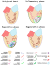
The phases of zebrafish cardiac regeneration. The uninjured heart contains a quiescent endocardium and epicardium, and a healthy myocardial layer. Following cardiac injury, the inflammatory phase takes place. Leucocytes are recruited to the injured area to remove necrotic and apoptotic cells and debris. Additionally, the endocardium and epicardium become activated and the first neovessels infiltrate the injury at the injury border zone (the interface between the injured myocardium and healthy myocardium). During the reparative phase, EPDC, synthetic mural cells and fibroblasts produce an extra-cellular matrix scaffold consisting of a collagen rich core and a fibrin cap. The dynamic signalling and activity of the epicardium and endocardium help coordinate the growth of new blood vessels and support the proliferation of existing cardiomyocytes at the injury border. Next, during the regenerative phase, proliferating cardiomyocytes repopulate the injured area and the fibrotic deposits are gradually removed. Finally, the endocardium and epicardium return to a quiescent state and the final maturation ensures the complete removal of scar tissue, restoration of cardiac function, and synchronicity. Figure created with BioRender.com.
Figure 4
Figure 4
Key signalling pathways in zebrafish endocardium and cardiac vessels during regeneration. Vessels at the injury border (orange circle) undergo a glycolytic switch and use glucose as a primary energy source. Border zone vessels express Apelin and Neuropilin 1 (Nrp1) and respond to Cxcl12b signals that originate at least in part from the activated epicardium. The activated endocardium (pink circle) expresses Notch, which regulates expression of endocardial maturation genes, endothelial differentiation, integrity, and pro-angiogenic genes (e.g., vegfc, klf2a/b). Additionally, endocardial Notch induces expression of Wnt antagonists wif1 and notum1b that in turn promote cardiomyocyte (CM) proliferation (Prolif). Activated endocardial cells express nrp1, aldh1a2, and runx1. A subpopulation of Runx1-positive cells expresses smooth muscle cell genes myh11a and taglnI. Runx1 also regulates anxa2 and serpine1 and limits fibrinolysis of scar tissue. Within the injury (blue circle), it is hypothesised that recruited macrophages and neutrophils combined with increased Hif1 activity upregulate Vegfa expression. An additional Vegfa source is thought to arise from proteolytic enzyme activity within the injury that releases extracellular matrix (ECM)–bound Vegfa. The activated epicardium (purple circle) secretes Cxcl12b chemokines that bind to the Cxcr4a receptor of endothelial cells in infiltrating vessels. Endothelial cell proliferation is further promoted by epicardial expression of Duox and Nox enzymes that catalyse the generation of hydrogen peroxide (H2O2). This subsequently inhibits Dusp6 activity in endothelial cells, thus relieving Dusp6 suppression of Erk signalling and enhancing endothelial proliferation. Fgf signalling promotes revascularisation and epicardial activation. Pdgf and Nrp1 promote epicardial activation, a subpopulation of epicardial cells undergo epicardial to mesenchymal transition (EpiMT). These epicardial-derived cells (EPDCs) become fibroblasts secreting ECM and perivascular cells. Nrg1 expression by EPDCs further promotes angiogenesis. Prox1a-expressing lymphatic vessels are detected in the injury and respond to Vegfc/d via Vegfr3. Inflammatory cells, necrotic and apoptotic cells, and extracellular matrix debris are removed via the lymphatic vessels (green circle) to aid the regenerative process. Figure generated with BioRender.com.

Similar articles

Cited by

References

    1. Kimmel C.B., Ballard W.W., Kimmel S.R., Ullmann B., Schilling T.F. Stages of embryonic development of the zebrafish. Dev. Dyn. 1995;203:253–310. doi: 10.1002/aja.1002030302. - DOI - PubMed
    1. Antinucci P., Hindges R. A crystal-clear zebrafish for in vivo imaging. Sci. Rep. 2016;6:29490. doi: 10.1038/srep29490. - DOI - PMC - PubMed
    1. Krauss J., Astrinidis P., Astrinides P., Frohnhöfer H.G., Walderich B., Nüsslein-Volhard C. Transparent, a gene affecting stripe formation in Zebrafish, encodes the mitochondrial protein Mpv17 that is required for iridophore survival. Biol. Open. 2013;2:703–710. doi: 10.1242/bio.20135132. - DOI - PMC - PubMed
    1. White A.J., Boffa M.B., Jones B., Petkovich M. A zebrafish retinoic acid receptor expressed in the regenerating caudal fin. Developement. 1994;120:1861–1872. - PubMed
    1. Isogai S., Horiguchi M., Weinstein B.M. The Vascular Anatomy of the Developing Zebrafish: An Atlas of Embryonic and Early Larval Development. Dev. Biol. 2001;230:278–301. doi: 10.1006/dbio.2000.9995. - DOI - PubMed

LinkOut - more resources