Review
. 2021 May 1;10(9):1954.
doi: 10.3390/jcm10091954.

Hypertrophic Cardiomyopathy and Primary Restrictive Cardiomyopathy: Similarities, Differences and Phenocopies

Affiliations
Review

Hypertrophic Cardiomyopathy and Primary Restrictive Cardiomyopathy: Similarities, Differences and Phenocopies

Riccardo Vio et al. J Clin Med. .

Abstract

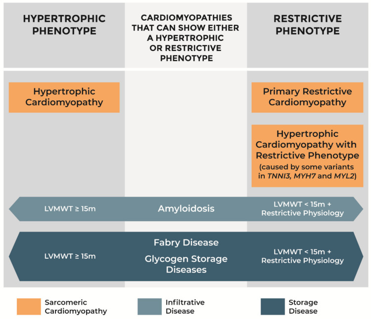
Hypertrophic cardiomyopathy (HCM) and primary restrictive cardiomyopathy (RCM) have a similar genetic background as they are both caused mainly by variants in sarcomeric genes. These "sarcomeric cardiomyopathies" also share diastolic dysfunction as the prevalent pathophysiological mechanism. Starting from the observation that patients with HCM and primary RCM may coexist in the same family, a characteristic pathophysiological profile of HCM with restrictive physiology has been recently described and supports the hypothesis that familiar forms of primary RCM may represent a part of the phenotypic spectrum of HCM rather than a different genetic cardiomyopathy. To further complicate this scenario some infiltrative (amyloidosis) and storage diseases (Fabry disease and glycogen storage diseases) may show either a hypertrophic or restrictive phenotype according to left ventricular wall thickness and filling pattern. Establishing a correct etiological diagnosis among HCM, primary RCM, and hypertrophic or restrictive phenocopies is of paramount importance for cascade family screening and therapy.

Keywords: Fabry disease; amyloidosis; cardiomyopathies; genetics; glycogen storage diseases; heart failure; hypertrophic cardiomyopathy; restrictive cardiomyopathy; restrictive physiology.


Conflict of interest statement

The authors declare no conflict of interest.

Figures

Figure 1
Figure 1
Restrictive form of heart failure due to diastolic dysfunction in a 28-year-old patient with non-obstructive hypertrophic cardiomyopathy preserved systolic function and troponin I variant. (A) Four-chamber view in end-diastole showing dilatation of both atria (left atrium, LA = 53 mm), normal-sized ventricles, and mild ventricular septal (VS) thickening (17 mm). (B) Pulsed Doppler waveform with evidence of restrictive filling: E/A>2; deceleration time, 150 ms. (C) Long-axis left ventricular (LV) plane with mild VS hypertrophy (17 mm); atria missing due to transplantation. (D,E) LV free wall (D) and septum (E) showing diffuse myocardial disarray, mild interstitial fibrosis, and intramural small vessel disease. Trichrome stain × 40. LVFW: left ventricular free wall; RA: right atrium; RV: right ventricle. Reproduced with permission from [29].
Figure 2
Figure 2
Novel Missense variant in MYL2 gene and hypertrophic cardiomyopathy associated with a high incidence of restrictive physiology. (A) Pedigree of the family. (B) Electrocardiogram of subject III-25 showing first-degree AV block and left anterior hemiblock (prior to pacemaker implantation). (C) Four-chamber echocardiographic view of subject III-25 showing biatrial enlargement and septal hypertrophy (prior to pacemaker implantation). (D) Gross view of the left cardiac chambers of subject III-25: note the severe dilatation of the left atrium with an almost preserved left ventricular volume. The thickness of the LV free wall and ventricular septum are 13 and 14 mm, respectively, in keeping with symmetric mild hypertrophy. (E) Histology of the LV free wall of subject III-25: note the diffuse disarray of the cardiac myocytes with tiny interstitial fibrosis (trichrome stain). Modified and reproduced with permission from [36].
Figure 3
Figure 3
Representative cardiac magnetic resonance findings of patients with obstructive hypertrophic cardiomyopathy, hypertrophic cardiomyopathy with restrictive phenotype, amyloidosis, and Fabry disease. (A) Obstructive HCM with intramural septal LGE. (B) HCM with restrictive physiology (MYH7 variant) with massive septal fibrosis and severe atrial enlargement. (C) Light chain immunoglobulin (AL) amyloidosis with transmural septal LGE. (D) Wild-type transthyretin (ATTRwt) amyloidosis with LGE particularly in the right ventricle and atria. (E) Fabry disease with hypertrophic phenotype and subendocardial LGE at the basal lateral segment of the left ventricle. (F) Fabry disease with mild hypertrophy and intramural LGE at the mid-lateral segment of the left ventricle and apex. HCM: hypertrophic cardiomyopathy; LGE: late gadolinium enhancement.
Figure 4
Figure 4
Phenotypes of hypertrophic cardiomyopathy, primary restrictive cardiomyopathy, amyloidosis, Fabry disease, and glycogen storage diseases. LVMWT: left ventricular maximal wall thickness.

References

    1. Maron B.J., Towbin J.A., Thiene G., Antzelevitch C., Corrado D., Arnett D., Moss A.J., Seidman C.E., Young J.B., American Heart Association et al. Contemporary definitions and classification of the cardiomyopathies: An American Heart Association Scientific Statement from the Council on Clinical Cardiology, Heart Failure and Transplantation Committee; Quality of Care and Outcomes Research and Functional Genomics and Translational Biology Interdisciplinary Working Groups; and Council on Epidemiology and Prevention. Circulation. 2006;113:1807–1816. - PubMed
    1. Ommen S.R., Mital S., Burke M.A., Day S.M., Deswal A., Elliott P., Evanovich L.L., Hung J., Joglar J.A., Kantor P., et al. 2020 AHA/ACC Guideline for the Diagnosis and Treatment of Patients with Hypertrophic Cardiomyopathy: A Report of the American College of Cardiology/American Heart Association Joint Committee on Clinical Practice Guidelines. Circulation. 2020;142:e558–e631. - PubMed
    1. Biagini E., Spirito P., Rocchi G., Ferlito M., Rosmini S., Lai F., Lorenzini M., Terzi F., Bacchi-Reggiani L., Boriani G., et al. Prognostic Implications of the Doppler Restrictive Filling Pattern in Hypertrophic Cardiomyopathy. Am. J. Cardiol. 2009;104:1727–1731. doi: 10.1016/j.amjcard.2009.07.057. - DOI - PubMed
    1. Mogensen J., Kubo T., Duque M., Uribe W., Shaw A., Murphy R., Gimeno J.R., Elliott P., McKenna W.J. Idiopathic restrictive cardiomyopathy is part of the clinical expression of cardiac troponin I mutations. J. Clin. Investig. 2003;111:209–216. doi: 10.1172/JCI200316336. - DOI - PMC - PubMed
    1. Angelini A., Calzolari V., Thiene G., Boffa G.M., Valente M., Daliento L., Basso C., Calabrese F., Razzolini R., Livi U., et al. Morphologic spectrum of primary restrictive cardiomyopathy. Am. J. Cardiol. 1997;80:1046–1050. doi: 10.1016/S0002-9149(97)00601-2. - DOI - PubMed

LinkOut - more resources