Review
. 2021 May 5;10(5):1107.
doi: 10.3390/cells10051107.

Fibrotic Events in the Progression of Cholestatic Liver Disease

Affiliations
Review

Fibrotic Events in the Progression of Cholestatic Liver Disease

Hanghang Wu et al. Cells. .

Abstract

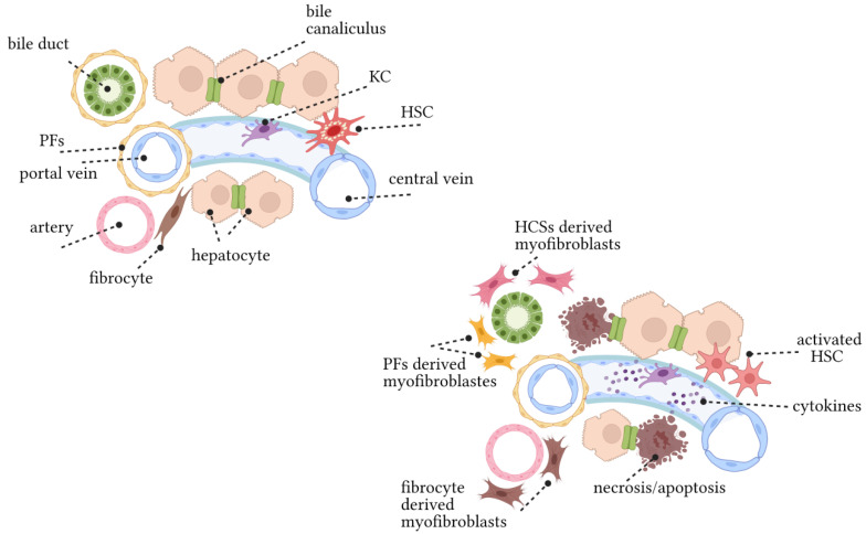
Cholestatic liver diseases including primary biliary cholangitis (PBC) and primary sclerosing cholangitis (PSC) are associated with active hepatic fibrogenesis, which can ultimately lead to the development of cirrhosis. However, the exact relationship between the development of liver fibrosis and the progression of cholestatic liver disease remains elusive. Periductular fibroblasts located around the bile ducts seem biologically different from hepatic stellate cells (HSCs). The fibrotic events in these clinical conditions appear to be related to complex crosstalk between immune/inflammatory mechanisms, cytokine signalling, and perturbed homeostasis between cholangiocytes and mesenchymal cells. Several animal models including bile duct ligation (BDL) and the Mdr2-knockout mice have improved our understanding of mechanisms underlying chronic cholestasis. In the present review, we aim to elucidate the mechanisms of fibrosis in order to help to identify potential diagnostic and therapeutic targets.

Keywords: cholangiocytes; cholestasis; fibrosis; hepatic stellate cells (HSCs); periductular fibroblasts.


Conflict of interest statement

The funders had no role in the design of the study; in the collection, analyses, or interpretation of data; in the writing of the manuscript, or in the decision to publish the results.

Figures

Figure 1
Figure 1
Pathogenesis of liver fibrosis. (Top left) The structure of liver lobules under physiological conditions. Biliary epithelial cells and hepatocytes, endothelial cells, HSCs, PFs, and KCs are the components of liver lobules. The bile duct, portal vein, and hepatic artery constitute the portal vein triad. HSCs are located in the space of Disse between hepatocytes and sinusoidal endothelium. HSCs are considered to be hepatic pericytes which contain lipid droplets and are the main storage cells for vitamin A. KCs are the resident liver macrophages. Only a few fibrocytes exist in a healthy liver. (Bottom right) Changes in liver lobules caused by chronic liver injury. In response to chronic liver injury, hepatocytes undergo apoptosis and release factors that recruit KCs, BM macrophages, and fibroblasts to sites of hepatic damage. KCs, macrophages, and fibroblasts release TGFβ1, which is one of the most powerful profibrogenic cytokines that can activate HSCs into collagen I-expressing myofibroblasts (e.g., αSMA). HSCs and PFs deposit ECM and a small number of fibroblasts.
Figure 2
Figure 2
Wnt/β-catenin, Notch, and Hedgehog signalling pathways. Wnt/β-catenin signalling is transmitted through the Frizzled (FZD) receptor, thereby stabilizing β-catenin. Phosphorylated β-catenin translocated into the nucleus to regulate the expression of target genes. There is crosstalk between Wnt/β-catenin signalling and Notch/TGF-β signalling. In Notch signalling, binding of Notch ligands to the receptor results in two proteolytic cleavages to release NICD. The released NICD then translocated into the nucleus, activating transcription factors Hes1, JAG1, and JAG2, whilst the Notch signal pathway interacts with Hh and Wnt signalling pathways. In Hedgehog signalling, Hh ligand secreted by Hedgehog secretory cells binds to PTCH or SMO and generates activated Gli that translocated to the nucleus, inducing target gene expression. These main survival pathways and sophisticated interactions between signalling pathways (i.e., TGF-β and MAPKs) constitute a complex regulatory network for the survival and proliferation of BECs.

References

    1. Erlinger S. What is cholestasis in 1985? J. Hepatol. 1985;1:687–693. doi: 10.1016/S0168-8278(85)80012-X. - DOI - PubMed
    1. Fuchs C.D., Halilbasic E., Trauner M. Pathophysiologic basis for alternative therapies for cholestasis. Liver Biol. Pathobiol. 2020:364–377. doi: 10.1002/9781119436812.ch30. - DOI
    1. Bataller R., Brenner D.A. Liver fibrosis. J. Clin. Investig. 2005;115:209–218. doi: 10.1172/JCI24282. - DOI - PMC - PubMed
    1. Kowdley K.V., Luketic V., Chapman R., Hirschfield G.M., Poupon R., Schramm C., Vincent C., Rust C., Pares A., Mason A., et al. A randomized trial of obeticholic acid monotherapy in patients with primary biliary cholangitis. Hepatology. 2018;67:1890–1902. doi: 10.1002/hep.29569. - DOI - PMC - PubMed
    1. Nevens F., Andreone P., Mazzella G., Strasser S.I., Bowlus C., Invernizzi P., Drenth J.P., Pockros P.J., Regula J., Beuers U., et al. A placebo-controlled trial of obeticholic acid in primary biliary cholangitis. N. Engl. J. Med. 2016;375:631–643. doi: 10.1056/NEJMoa1509840. - DOI - PubMed

Publication types

LinkOut - more resources