. 2021 May 5;22(9):4877.
doi: 10.3390/ijms22094877.

Molecular, Cellular and Functional Changes in the Retinas of Young Adult Mice Lacking the Voltage-Gated K+ Channel Subunits Kv8.2 and K2.1

Affiliations

Molecular, Cellular and Functional Changes in the Retinas of Young Adult Mice Lacking the Voltage-Gated K+ Channel Subunits Kv8.2 and K2.1

Xiaotian Jiang et al. Int J Mol Sci. .

Abstract

Cone Dystrophy with Supernormal Rod Response (CDSRR) is a rare autosomal recessive disorder leading to severe visual impairment in humans, but little is known about its unique pathophysiology. We have previously shown that CDSRR is caused by mutations in the KCNV2 (Potassium Voltage-Gated Channel Modifier Subfamily V Member 2) gene encoding the Kv8.2 subunit, a modulatory subunit of voltage-gated potassium (Kv) channels. In a recent study, we validated a novel mouse model of Kv8.2 deficiency at a late stage of the disease and showed that it replicates the human electroretinogram (ERG) phenotype. In this current study, we focused our investigation on young adult retinas to look for early markers of disease and evaluate their effect on retinal morphology, electrophysiology and immune response in both the Kv8.2 knockout (KO) mouse and in the Kv2.1 KO mouse, the obligate partner of Kv8.2 in functional retinal Kv channels. By evaluating the severity of retinal dystrophy in these KO models, we demonstrated that retinas of Kv KO mice have significantly higher apoptotic cells, a thinner outer nuclear cell layer and increased activated microglia cells in the subretinal space. Our results indicate that in the murine retina, the loss of Kv8.2 subunits contributes to early cellular and physiological changes leading to retinal dysfunction. These results could have potential implications in the early management of CDSRR despite its relatively nonprogressive nature in humans.

Keywords: CDSRR; KCNB1; KCNV2; cone-rod dystrophy; photoreceptors; retinal degeneration; voltage-gated potassium channels.


Conflict of interest statement

The authors declare no conflict of interest. The funders had no role in the design of the study; in the collection, analyses, or interpretation of data; in the writing of the manuscript, or in the decision to publish the results.

Figures

Figure 1
Figure 1
Retinal localization and expression of the voltage-gated K+ channel protein subunits Kv8.2 and Kv2.1 in wild type (WT), Kv8.2 knockout (KO) and Kv2.1 KO. (AC) Representative confocal images of the inner segment (IS) area, outer nuclear layer (ONL), inner nuclear layer (INL) and ganglion cell layer (GCL) showing expression of Kv8.2 and Kv2.1 mouse subunits in wild type (A), Kv8.2 KO (B) and Kv2.1 KO (C) retinas at 20× (top panels) and 40× (bottom panels) magnification. Scale bars = 20 μM (top panels) and 50 μM (bottom panels). (D,E) Relative mRNA expression of Kcnv2 and Kcnb1 genes in all three lines. Results are presented as mean +/− SD from n = 3 (WT and Kv8.2 KO) and n = 4 (Kv2.1 KO), and p values were obtained through one-way ANOVA and Dunnett’s multiple comparison test post hoc with * p = 0.0094 and ** p = 0.0001.
Figure 2
Figure 2
Representative confocal images of wild type and knockout (KO) retinas from central (10° from optic nerve) dorsal-ventral areas showing signal overlap of (A) rhodopsin (Rho, red) or (B) cone arrestin (Arr3, red) with both Kv subunits in wild type and Kv2.1 subunit in Kv8.2 KO retinas (green). No anti-Kv8.2 and anti-Kv2.1 staining was observed in the Kv2.1 KO retinas. ONL, outer nuclear layer; IS, inner segment; INL, inner nuclear layer; OS, outer segment. Scale bar = 20 μM.
Figure 3
Figure 3
Retinal thickness and cell death in wild type (WT) and knockout (KO) retinas. (A) Representative histological retinal sections from the dorsal-ventral region of KO models and WT showing the different retinal layers. RPE, retinal pigment epithelium; OS/IS, outer segment/inner segment; ONL, outer nuclear layer; OPL, outer plexiform layer; INL, inner nuclear layer; IPL, inner plexiform layer; RGC, retinal ganglion cells. Scale bar = 100 μM. (B) Quantification of outer nuclear layer (ONL) thickness in the different mouse models in central (10° from optic nerve) and peripheral (80° from optic nerve) areas of the retina. No differences were observed between dorsal or ventral areas. The results are presented as mean +/− SEM, with statistical analysis by two-way ANOVA and Sidak’s multiple comparison test post hoc with * p < 0.002. (C) Quantification of cell death in the ONL of WT, Kv8.2 KO and Kv2.1 KO retinas via TUNEL (Terminal deoxynucleotidyl transferase dUTP Nick End Labeling) staining. Results are presented as mean +/− SD from n = 3–5, and p values were obtained through one-way ANOVA and Tukey’s multiple comparison test post hoc with * p < 0.006.
Figure 4
Figure 4
Glia activation in Kv deficient retinas. (A) Glial fibrillary acidic protein (GFAP) retinal expression in wild type, Kv8.2 KO and Kv2.1 KO animals. Upper panels show nuclear stain (DAPI, blue) and GFAP (red) co-localisation. Boomt panels show GFAP (red) expression only. Scale bar = 50 μM (B) Fluorescent quantification of GFAP protein expression in the three mouse lines showing significantly higher expression in both Kv KO lines compared to WT. (C) Quantification of Gfap gene expression via qPCR showing a significant increase in expression in Kv2.1 KO retinas compared to wild type (n = 3 for each genotype). (D) Confocal images of retinal flatmounts labeled with microglia marker Iba-1 (red) taken at the inner nuclear layer (INL) and outer nuclear layer (ONL) regions to show proliferation and migration of activated microglia in the Kv8.2 KO retina. Scale bar = 50 μM. (E) Higher magnification images of individual microglia cells from WT (i), Kv8.2 KO INL (ii) and ONL (iii), and Kv2.1 KO INL (iv) showing the morphological differences in cell body shape. (F) Quantification of microglia numbers in the INL and ONL in the wild type, Kv8.2 KO and Kv2.1 KO retinas. Results are presented as mean +/− SD from n = 3, and p values were obtained through two-way ANOVA and Tukey’s multiple comparison test post hoc with * p < 0.02, ** p < 0.004, *** p < 0.0006, **** p < 0.0001.
Figure 5
Figure 5
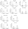
Characterization of immune cells present in the retina of wild type (WT) and Kv8.2 knockout (KO) mice. Panels show quantification of different types of immune cells in two-month-old WT and Kv8.2 KO retinas. Results are presented as mean +/− SEM from n = 6. p values were obtained through unpaired t-test with Welch’s correction, * p = 00286, ** p < 0.0029 and *** p = 0.0002.
Figure 6
Figure 6
Photopic visual response from Kv deficient retinas. (A) Representative light-adapted photopic full flash ERG traces from wild type (WT), Kv8.2 knockout (KO) and Kv2.1 KO animals taken at 25 cd.s/m2. (B) Quantification of photopic a-wave in all three genotypes at different stimulus intensities. * denote significance between the Kv lines and WT with p < 0.0005. No statistical difference was observed between the Kv8.2 and Kv2.1 KO lines. (C) Quantification of photopic b-wave in all three genotypes at different stimulus intensities. * denote significance between the Kv lines and WT with p < 0.0005. No statistical difference was observed between the Kv8.2 and Kv2.1 KO lines. (D) Light-adapted photopic optomotor response from all three genotypes. Y-axis shows tracking movements per 2 min. * p < 0.002 compared to WT. Results for (BD) are presented as mean +/− SD from n = 3–4 animals (6 eyes per genotype). Significance was obtained through a two-way ANOVA and Tukey’s multiple comparison test post hoc.
Figure 7
Figure 7
Scotopic ERG response. (A) Representative traces of dark-adapted full flash scotopic responses at 25 cd.s/m2. Traces show the distinctive supernormal b-wave (+b) component of the ERG trace in both Kv8.2 and Kv2.1 KO animals. Amplitude length of a-wave (a) and full b-wave (b) are also shown. (B) Quantification of the dark-adapted scotopic a-wave, b-wave and positive b-wave components at different light intensities for WT, Kv8.2 KO and Kv2.1 KO animals. Results are presented as mean +/− SD from n = 3–4 animals (six eyes per genotype); significance was obtained through a two-way ANOVA and Tukey’s multiple comparison test post hoc. a-wave: * p < 0.0001 at all intensities for both Kv lines compared to WT; ** p = 0.0085 between Kv8.2 and Kv2.1 KO lines. b-wave: * p < 0.01 between Kv8.2 and Kv2.1 KO lines only. Not significant compared to WT. Positive b-wave: ** p = 0.0025 only between WT and Kv8.2 only. * p = 0.004 (0.3 cd.s m−2) and p < 0.0006 (1–25 cd.s m−2) between Kv lines and WT. Not significant between Kv lines.

Similar articles

Cited by

References

    1. Gouras P., Eggers H.M., Mackay C.J. Cone Dystrophy, Nyctalopia, and Supernormal Rod Responses. A new retinal degeneration. Arch. Ophthalmol. 1983;101:718–724. doi: 10.1001/archopht.1983.01040010718003. - DOI - PubMed
    1. Wu H., Cowing J.A., Michaelides M., Wilkie S.E., Jeffery G., Jenkins S.A., Mester V., Bird A.C., Robson A.G., Holder G.E., et al. Mutations in the Gene KCNV2 Encoding a Voltage-Gated Potassium Channel Subunit Cause “Cone Dystrophy with Supernormal Rod Electroretinogram” in Humans. Am. J. Hum. Genet. 2006;79:574–579. doi: 10.1086/507568. - DOI - PMC - PubMed
    1. Wissinger B., Dangel S., Jagle H., Hansen L., Baumann B., Rudolph G., Wolf C., Bonin M., Koeppen K., Ladewig T., et al. Cone Dystrophy with Supernormal Rod Response Is Strictly Associated with Mutations in KCNV2. Investig. Opthalmol. Vis. Sci. 2008;49:751–757. doi: 10.1167/iovs.07-0471. - DOI - PubMed
    1. Wissinger B., Schaich S., Baumann B., Bonin M., Jägle H., Friedburg C., Varsányi B., Hoyng C.B., Dollfus H., Heckenlively J.R., et al. Large deletions of the KCNV2 gene are common in patients with cone dystrophy with supernormal rod response. Hum. Mutat. 2011;32:1398–1406. doi: 10.1002/humu.21580. - DOI - PubMed
    1. Zelinger L., Wissinger B., Eli D., Kohl S., Sharon D., Banin E. Cone Dystrophy with Supernormal Rod Response: Novel KCNV2 Mutations in an Underdiagnosed Phenotype. Ophthalmology. 2013;120:2338–2343. doi: 10.1016/j.ophtha.2013.03.031. - DOI - PubMed