Review
. 2021 May 5;22(9):4892.
doi: 10.3390/ijms22094892.

Preventive Applications of Polyphenols in Dentistry-A Review

Affiliations
Review

Preventive Applications of Polyphenols in Dentistry-A Review

Jasmin Flemming et al. Int J Mol Sci. .

Abstract

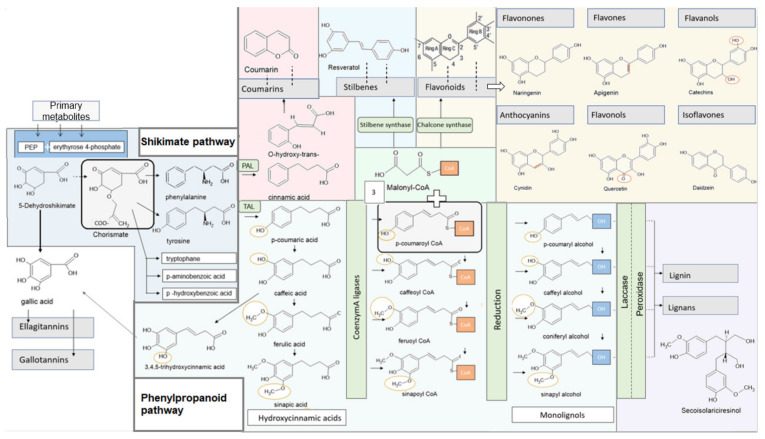
Polyphenols are natural substances that have been shown to provide various health benefits. Antioxidant, anti-inflammatory, and anti-carcinogenic effects have been described. At the same time, they inhibit the actions of bacteria, viruses, and fungi. Thus, studies have also examined their effects within the oral cavity. This review provides an overview on the different polyphenols, and their structure and interactions with the tooth surface and the pellicle. In particular, the effects of various tea polyphenols on bioadhesion and erosion have been reviewed. The current research confirms that polyphenols can reduce the growth of cariogenic bacteria. Furthermore, they can decrease the adherence of bacteria to the tooth surface and improve the erosion-protective properties of the acquired enamel pellicle. Tea polyphenols, especially, have the potential to contribute to an oral health-related diet. However, in vitro studies have mainly been conducted. In situ studies and clinical studies need to be extended and supplemented in order to significantly contribute to additive prevention measures in caries prophylaxis.

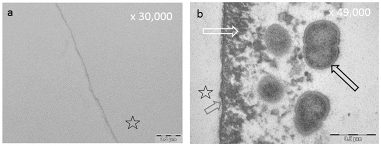
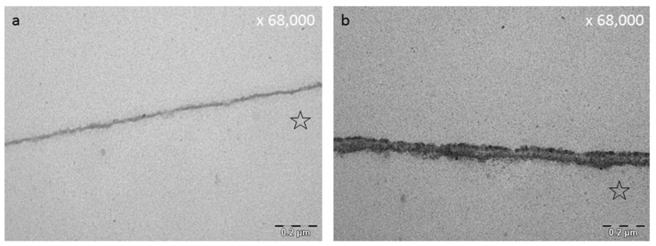
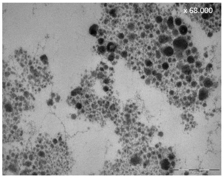
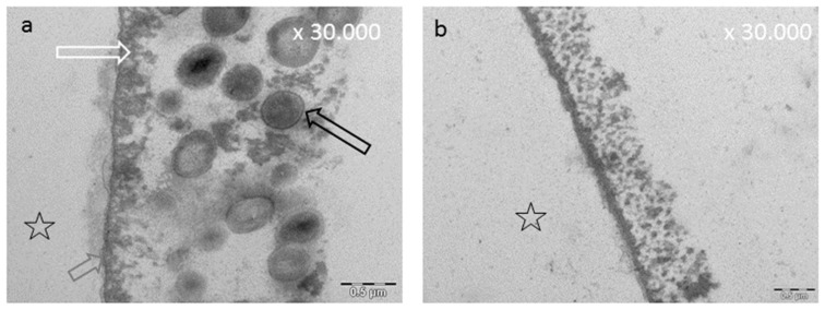
Keywords: medicinal plants; molecular mechanisms; polyphenols; preventive dentistry; salivary pellicle; tea drugs; transmission electron microscopy (TEM).


Conflict of interest statement

The authors declare that they have no competing interests.

Figures

Figure 1
Figure 1
Illustration of phenylpropanoids synthesis and the synthesis of various polyphenols.
Figure 2
Figure 2
Illustration of the delocalization of electrons.
Figure 3
Figure 3
Representative TEM images of the 10 s (a) and the 24 h pellicle layer (b). The formation of a dense basal layer (b, grey arrow) can already be visualized after a few seconds. After 2 h, there are granular and globular structures (b, white arrow) which have absorbed onto this basal layer due to protein–protein interactions. As a result, an increase in pellicle thickness can be observed. (b) The result after 24 h biofilm formation. Please note the segmentation of the bacterial cell, indicating viability of the microorganisms (b, black arrow). Please note that all TEM images (Figure 3, Figure 4, Figure 5, Figure 7, Figure 8 and Figure 10) depict pellicles formed in situ. For this purpose, bovine enamel slabs were fixed to individual splints with silicone impression material for defined exposure to the oral fluids. The enamel was removed during the extensive sample preparation by acid etching; the former enamel site is marked with an asterisk. For details of the methods, please refer to Hertel et al. (2016) [77], Rehage et al. (2017) [68], and Kirsch et al. (2020) [149].
Figure 4
Figure 4
Effect of polyphenols on the pellicle’s ultrastructure, representative TEM images of the in situ pellicle after rinsing with tannic acid—oral exposure time 30 min: the control (a) shows a thin basal pellicle layer (Rehage et al. (2017) [68]; (b) a distinct increase in pellicle thickness and electron density after ex vivo rinsing with tannic acid (170 mg/mL, pH = 5.0). The former enamel site is marked with an asterisk.
Figure 5
Figure 5
Both TEM images visualize the 2 h pellicle after a pellicle formation time of 1 min and rinsing with tannic acid for 10 min (a,b). (b) Sample generated after an additional treatment with HCl (pH 2.3) for 1 min in vitro (Hertel et al. (2017) [177]. In comparison, Figure 7a shows a 2 h control pellicle without rinsing. The former enamel site is marked with an asterisk.
Figure 6
Figure 6
Denaturation of pellicle proteins through polyphenols. (a) Simplified representation of the pellicle ultrastructure on the tooth surface with an attached three-dimensional biofilm. Thereby, some proteins serve as receptors for bacterial cells to adhere to the tooth surface. (b) Hampered bacterial adhesion at the tooth surface. Polyphenols cause denaturation and changes in secondary and tertiary structure of proteins. In the following binding of bacteria to the polyphenol-modified-receptors is inhibited. Polyphenols also lead to a lysis of bacterial cells. In addition, polyphenol-protein aggregates adhere at the tooth surface. Consequently, the pellicle is denser and thicker in its ultrastructure.
Figure 7
Figure 7
Representative TEM images of the pellicle ultrastructure after initial rinsing with different polyphenolic agents followed by 2 h of intraoral exposure. Rinsing with polyphenolic compounds leads to an increased pellicle thickness and density. The former enamel site was marked with an asterisk. (a) Native pellicle, control, no rinsing, intraoral exposure: 2 h, (b) Control, effect of CHX, intraoral exposure: 2 h, (c) Leaves of R. nigrum, watery plant extract 8 mL, 10 min rinsing, intraoral exposure: 2 h. For details consider Weber et al. (2015) [178], (d) Hamamelis (bark), 0.2 mg/8 mL, 10 min rinsing, intraoral exposure: 2 h. For details consider Kirsch et al. (2020) [149].
Figure 8
Figure 8
TEM images visualizing the effect of cistus incanus tea on the in situ pellicle layer after rinsing with 8 mL of cistus incanus tea for 10 min (Wittpahl et al. (2015) [63]). (a) shows the results after 30 min, (b) after 120 min. The former enamel site was marked with an asterisk.
Figure 9
Figure 9
Effect of polyphenols on salivary proteins: Representative TEM-image of a saliva-sample after 30-s in vivo rinsing with 5% tannic acid, pH 2.0. The rinsing of tannic acid leads to aggregation of polyphenolic compounds with salivary proteins visible as electron dense globules.
Figure 10
Figure 10
Sustainable long-term effects of tannic acid on bioadhesion and biofilm formation in situ, TEM. After 24 h of oral exposure, bacterial biofilm formation was observed (a,b) as also described in Xi et al. (2020) [259]. (a) shows an image of the control without rinsing. The basal pellicle layer is marked with a grey arrow, the globular pellicle layer is marked with a white arrow and the bacterial cell is marked with a black arrow. Tannic acid modified pellicle is covered by bacteria and extracellular biofilm matrix (b). The volunteers rinsed with a solution containing 1% tannic acid and 1% chinese gallnut extract, 2 times a day. The first time 3 min after the splint was inserted, the second time before night sleeping. As also seen in Figure 9, aggregates enriched from the saliva can be observed above the pellicle. The former enamel site was marked with an asterisk.

References

    1. Unachukwu U.J., Ahmed S., Kavalier A., Lyles J.T., Kennelly E.J. White and Green Teas (Camellia sinensis var. sinensis): Variation in Phenolic, Methylxanthine, and Antioxidant Profiles. J. Food Sci. 2010;75:C541–C548. doi: 10.1111/j.1750-3841.2010.01705.x. - DOI - PubMed
    1. Ditano-Vazquez P., Torres-Pena J.D., Galeano-Valle F., Perez-Caballero A.I., Demelo-Rodriguez P., Lopez-Miranda J., Katsiki N., Delgado-Lista J., Alvarez-Sala-Walther L.A. The Fluid Aspect of the Mediterranean Diet in the Prevention and Management of Cardiovascular Disease and Diabetes: The Role of Polyphenol Content in Moderate Consumption of Wine and Olive Oil. Nutrients. 2019;11:2833. doi: 10.3390/nu11112833. - DOI - PMC - PubMed
    1. Niemetz R., Gross G.G. Enzymology of gallotannin and ellagitannin biosynthesis. Phytochemistry. 2005;66:2001–2011. doi: 10.1016/j.phytochem.2005.01.009. - DOI - PubMed
    1. Hannig C., Attin T., Hannig M., Henze E., Brinkmann K., Zech R. Immobilisation and activity of human alpha-amylase in the acquired enamel pellicle. Arch. Oral Biol. 2004;49:469–475. doi: 10.1016/j.archoralbio.2004.01.005. - DOI - PubMed
    1. He J., Li Y., Cao Y., Xue J., Zhou X. The oral microbiome diversity and its relation to human diseases. Folia Microbiol. 2015;60:69–80. doi: 10.1007/s12223-014-0342-2. - DOI - PubMed

LinkOut - more resources