

Post-Surgical Peritoneal Scarring and Key Molecular Mechanisms
- PMID: 34063089
- PMCID: PMC8147932
- DOI: 10.3390/biom11050692
Post-Surgical Peritoneal Scarring and Key Molecular Mechanisms
Abstract
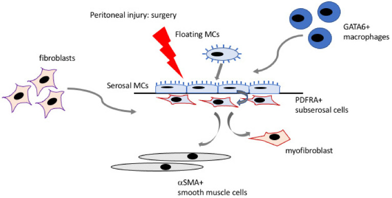
Post-surgical adhesions are internal scar tissue and a major health and economic burden. Adhesions affect and involve the peritoneal lining of the abdominal cavity, which consists of a continuous mesothelial covering of the cavity wall and majority of internal organs. Our understanding of the full pathophysiology of adhesion formation is limited by the fact that the mechanisms regulating normal serosal repair and regeneration of the mesothelial layer are still being elucidated. Emerging evidence suggests that mesothelial cells do not simply form a passive barrier but perform a wide range of important regulatory functions including maintaining a healthy peritoneal homeostasis as well as orchestrating events leading to normal repair or pathological outcomes following injury. Here, we summarise recent advances in our understanding of serosal repair and adhesion formation with an emphasis on molecular mechanisms and novel gene expression signatures associated with these processes. We discuss changes in mesothelial biomolecular marker expression during peritoneal development, which may help, in part, to explain findings in adults from lineage tracing studies using experimental adhesion models. Lastly, we highlight examples of where local tissue specialisation may determine a particular response of peritoneal cells to injury.
Keywords: biomarkers; mesothelium; molecular signatures; peritoneum; post-surgical adhesions; serosal repair.
Conflict of interest statement
The authors declare no conflict of interest.
Figures
References
Publication types
MeSH terms
Substances
Grants and funding
LinkOut - more resources
Full Text Sources
Medical
