Review
. 2021 May 2;13(5):824.
doi: 10.3390/v13050824.

Complement Proteins as Soluble Pattern Recognition Receptors for Pathogenic Viruses

Affiliations
Review

Complement Proteins as Soluble Pattern Recognition Receptors for Pathogenic Viruses

Valarmathy Murugaiah et al. Viruses. .

Abstract

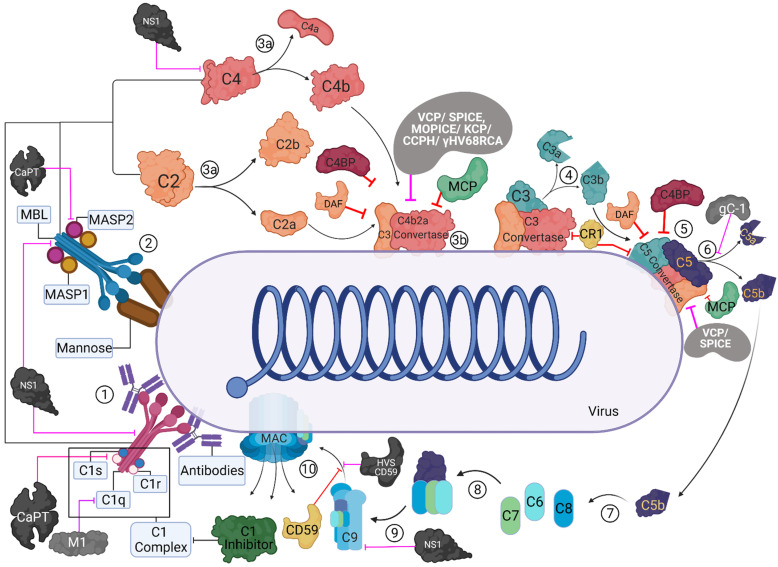
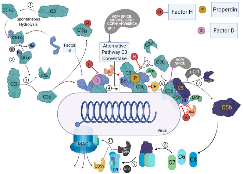
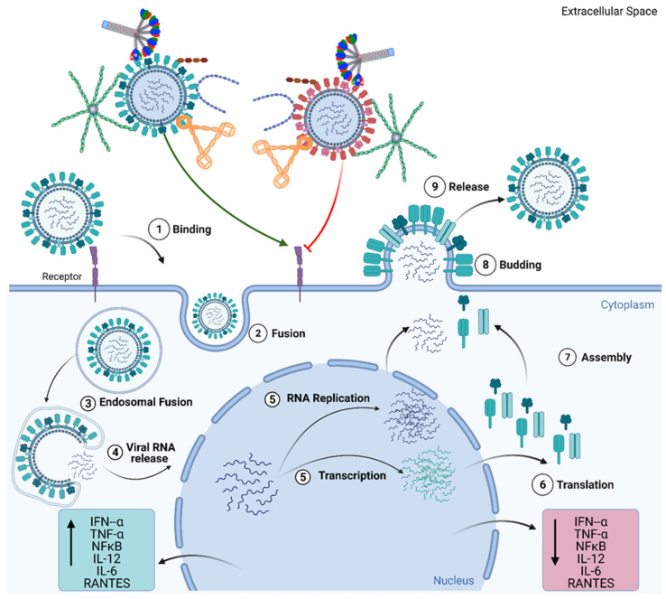
The complement system represents a crucial part of innate immunity. It contains a diverse range of soluble activators, membrane-bound receptors, and regulators. Its principal function is to eliminate pathogens via activation of three distinct pathways: classical, alternative, and lectin. In the case of viruses, the complement activation results in effector functions such as virion opsonisation by complement components, phagocytosis induction, virolysis by the membrane attack complex, and promotion of immune responses through anaphylatoxins and chemotactic factors. Recent studies have shown that the addition of individual complement components can neutralise viruses without requiring the activation of the complement cascade. While the complement-mediated effector functions can neutralise a diverse range of viruses, numerous viruses have evolved mechanisms to subvert complement recognition/activation by encoding several proteins that inhibit the complement system, contributing to viral survival and pathogenesis. This review focuses on these complement-dependent and -independent interactions of complement components (especially C1q, C4b-binding protein, properdin, factor H, Mannose-binding lectin, and Ficolins) with several viruses and their consequences.

Keywords: DNA viruses; RNA viruses; complement evasion; complement system; cytokine storm; innate immunity; retroviruses.


Conflict of interest statement

The authors declare no conflict of interest.

Figures

Figure 1
Figure 1
Activation and regulation of the classical and lectin pathways and their targeting by virally encoded molecules. In the classical pathway (CP), C1 complex recognizes the antigen-antibody complexes present on the viral surface (1). In the lectin pathway (LP), MBL/ficolin-MASP complexes can recognise other carbohydrate patterns on the surfaces of viruses (2). Upon activation, these complexes can cleave C4 and C2 (3a) that can lead to the formation of C4bC2a (CP/LP C3 convertase) (3b). The C3 convertase further cleaves C3 into C3b and C3a; C3b is known to opsonise the viral surfaces, whereas C3a can lead to an enhanced acquired immune responses (4). C3b-C3 convertase interaction can generate C5 convertase (5), which cleaves C5 into C5b and C5a (6). C5b further interacts with C6 and C7 (C5b-7) (7) that can bind to the viral surface, while C5a induces further infiltration. C5b-7 then binds to C8, which can generate C5b-8 that penetrates the membrane (8). Finally, the C9 binds to the C5b-8 and results in MAC formation leading to the virolysis (10). These activation pathways are regulated at different steps by host complement regulators such as C1 inhibitor, C4b-binding protein (C4BP), complement receptor 1 (CR1; CD35), membrane cofactor protein (MCP; CD46), decay-accelerating factor (DAF; CD55), and CD59. Viral proteins that target these pathways are: Vaccinia virus complement control protein (VCP), Smallpox inhibitor of complement enzymes (SPICE), Monkeypox inhibitor of complement enzymes (MOPICE), Kaposi’s sarcoma-associated herpesvirus inhibitor of complement activation (KCP), Murine gamma-herpesvirus 68 regulator of complement activation (γ-HV68 RCA), Herpesvirus saimiri complement control protein homologue (CCPH), Herpesvirus saimiri CD59 homologue (HVS CD59), Flavivirus non-structural protein 1 (NS1), HSV-1 glycoprotein C (gC-1), human astrovirus coat protein (CoPt), and Influenza virus matrix protein 1 (M1). These are identified as black/grey protein with white text, and pink inhibitory arrows mark the regulator they inhibit.
Figure 2
Figure 2
The activation and regulation of the alternative pathway and its targeting by virally encoded molecules. During the process of alternative pathway (AP), native C3 by H2O is spontaneously hydrolysed, resulting in the formation of C3b like C3 [C3(H2O)] (1). C3(H2O) can bind to factor B (FB), and upon cleavage by factor D (FD), which forms the initial AP-derived C3 convertase (2). The C3 convertase can cleave C3 into C3b and C3a (3). The C3b then binds to the viral surfaces, and trigger the formation of surface bound C3bBb, with the involvement of FB and FD (4). The surface bound C3bBb can then initiate the amplification loop of the AP (5), causing deposition of C3b molecules on to viral surfaces. C3b can combine with pre-existing AP-derived C3 convertase, which leads to the formation of C5 convertase (6). C5 convertase cleaves C5 into C5b and C5a (7). C5b further interacts with C6 and C7 to form C5b-7 (8), which can bind to the surfaces of viruses, while C5a acts as an anaphylatoxins. C5b-7 then binds to C8 which can generate C5b-8 that penetrates the membrane (9). Finally, the C9 binds to C5b-8, resulting in MAC formation (10). The activation steps are regulated at different steps by host complement regulators such as complement receptor 1 (CR1; CD35), membrane cofactor protein (MCP, CD46), decay-accelerating factor (DAF; CD55), factor H (FH), and CD59. Viral proteins that target these pathways are: Vaccinia virus complement control protein (VCP), Smallpox inhibitor of complement enzymes (SPICE), Monkeypox inhibitor of complement enzymes (MOPICE), Kaposi’s sarcoma-associated herpesvirus inhibitor of complement activation (KCP), Murine gamma-herpesvirus 68 regulator of complement activation (γ-HV68 RCA), Herpesvirus saimiri complement control protein homologue (CCPH), Herpesvirus saimiri CD59 homologue (HVS CD59), Flavivirus non-structural protein 1 (NS1), and HSV-1 glycoprotein C (gC-1). These viral proteins are identified as black/grey proteins with white text, and pink inhibitory arrows mark the regulator they inhibit.
Figure 3
Figure 3
Complement Independent functions of Complement Regulators. Viral infection begins with the attachment of the virus to the epithelial cell surface via cell surface receptors (1) and the internalisation of the virion through endocytosis and fusion (2). Post endocytosis, viral RNA is released into the cytoplasm (3,4), from where it is transported into the nucleus. In the nucleus, the viral RNA undergoes replication and transcription (5). The transcribed mRNA is translated to viral proteins (6). This is followed by the assembly of the virion and subsequent release of the virion from the cell (7,8,9). C1q, C4BP, Properdin, factor H, and VCP have individually been shown to inhibit the entry of viruses, such as the H1N1 subtype of the Influenza A Virus (IAV), (represented by red virion) into the cell and downregulate inflammatory cytokines and chemokines (TNF-α, IL-6, IL-12, NF-κB, RANTES). However, these complement regulators individually have also been implicated in promoting viral entry, as seen in the case of H3N2 subtype of IAV, and promoting the inflammatory response by upregulating cytokine and chemokines (TNF-α, IL-6, IL-12, NF-κB, and RANTES). These mechanisms of modulating viral entry in a subtype specific manner, occur in the absence of other complement factors and immune cells, suggesting complement independent viral infection modulating activity for these complement regulatory proteins.

References

    1. Sim R.B., Tsiftsoglou S.A. Proteases of the complement system. Biochem. Soc. Trans. 2004;32:21–27. doi: 10.1042/bst0320021. - DOI - PubMed
    1. Müller-Eberhard H.J., Polley M.J., Calcott M.A. Formation and Functional Significance of a Molecular Complex Derived from the Second and the Fourth Component of Human Complement. J. Exp. Med. 1967;125:359–380. doi: 10.1084/jem.125.2.359. - DOI - PMC - PubMed
    1. Naff G.B., Pensky J., Lepow I.H. The Macromolecular Nature of the First Component of Human Complement. J. Exp. Med. 1964;119:593–613. doi: 10.1084/jem.119.4.593. - DOI - PMC - PubMed
    1. Polley M.J., Müller-Eberhard H.J. The second component of human complement: Its isolation, fragmentation by C’1 esterase, and incorporation into C’3 convertase. J. Exp. Med. 1968;128:533–551. doi: 10.1084/jem.128.3.533. - DOI - PMC - PubMed
    1. Kishore U., Reid K.B. Modular organization of proteins containing C1q-like globular domain. Immunopharmacology. 1999;42:15–21. doi: 10.1016/S0162-3109(99)00011-9. - DOI - PubMed

MeSH terms

Substances

LinkOut - more resources