Review
. 2021 May 15;26(10):2940.
doi: 10.3390/molecules26102940.

Electrochemical Biosensors in Food Safety: Challenges and Perspectives

Affiliations
Review

Electrochemical Biosensors in Food Safety: Challenges and Perspectives

Antonella Curulli. Molecules. .

Abstract

Safety and quality are key issues for the food industry. Consequently, there is growing demand to preserve the food chain and products against substances toxic, harmful to human health, such as contaminants, allergens, toxins, or pathogens. For this reason, it is mandatory to develop highly sensitive, reliable, rapid, and cost-effective sensing systems/devices, such as electrochemical sensors/biosensors. Generally, conventional techniques are limited by long analyses, expensive and complex procedures, and skilled personnel. Therefore, developing performant electrochemical biosensors can significantly support the screening of food chains and products. Here, we report some of the recent developments in this area and analyze the contributions produced by electrochemical biosensors in food screening and their challenges.

Keywords: antibiotics; bacteria; contaminants; electrochemical biosensors; food; pesticides; safety; toxins.


Conflict of interest statement

The author declares no conflict of interest.

Figures

Figure 1
Figure 1
Classification of biosensors based on various bioreceptors and transducers used [16].
Figure 2
Figure 2
Scheme of a biosensor with an electrochemical transducer. Reprinted with permission from [10] Copyright (2010) Royal Society of Chemistry (RSC).
Figure 3
Figure 3
Schematic diagram of (a) amperometric/voltammetric, (b) potentiometric, (c) conductometric biosensors, and (d) impedimetric biosensor with the relative equivalent circuit [16] (Cdl = double-layer capacitance of the electrodes, Rsol = resistance of the solution, Cde = capacitance of the electrode, Zcell = impedance introduced by the bound nanoparticles, and Rcell and Ccell are the resistance and capacitance in parallel).
Figure 4
Figure 4
Schematic diagram of the M-GO-assisted homogeneous ECL aptasensor for OA determination. Reprinted with permission from [57] Copyright 2021 Elsevier.
Figure 5
Figure 5
Schematic representation related to the ECL Escherichia coli biosensor, fabricated with luminol/AgBr/3DNGH. Reprinted with permission from [116] Copyright 2017 Elsevier.
Figure 6
Figure 6
Schematic representation of the assembling and the sensing approach of the E. coli aptasensor. Reprinted with permission from [117] Copyright 2017 Elsevier.
Figure 7
Figure 7
Schematic representation of the biosensor for S. aureus based on a DNA walker and DNA nanoflowers. Reprinted with permission from [126] Copyright 2021 American Chemical Society.
Figure 8
Figure 8
Schematic diagram of the synthesis of MXene nanosheets and assembling of the enzyme-based pesticide biosensor. Reprinted with permission from [150] Copyright 2020 Elsevier.
Figure 9
Figure 9
(A) Cyclic voltammograms, (B) differential pulse voltammograms and (C) square wave voltammograms of Ag-citrate/GQDs nano-ink fabricated on the surface of apple-skin incubated at room temperature in the absence and presence of 1 mM trifluralin. Supporting electrolyte is 0.1 M PBS (pH 7.4) in the presence of acetone, (D) photographic image of an electrochemical sensor made by direct writing of nano-ink on the surface of apple skin. Reprinted with permission from [158] Copyright 2020 Elsevier.
Figure 10
Figure 10
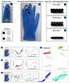
(A) Details of finger sensor design with a complete electrochemical system: auxiliary, reference and working electrodes. The connection between electrodes and potentiostat was made via flexible conductive wires for on-site detection. (B) Image of the real screen-printed sensing glove. (CE) Schematic representation of the side views of CSS, PCNB and pretreated sensing layers for index, middle and ring fingers, respectively. The electrochemical signatures and corresponding analytical curves obtained with index, middle and ring fingers of the glove-embedded sensors are shown in (F) through (H,F) DPV for carbendazim detection from 1.0 × 10⁻⁷ to 1.0 × 10⁻⁶ mol L–1; (G) DPV for diuron detection from 1.0 × 10⁻⁷ to 1.0 × 10⁻⁶ mol L–1; (H) SW voltammograms for paraquat detection from 1.0× from 1.0 × 10⁻⁷ to 1.0 × 10⁻⁶ mol L−1 and fenitrothion detection from 1.0 × 10⁻⁷ to 1.0 × 10⁻⁶ mol L−1. Conditions for the detection: 0.1 mol L−1 phosphate buffer solution, pH 7.0. (I) LSP plot for all pesticides measured with differential pulse and square wave voltammetry, where each voltammogram was converted into a colored dot on the plot. The black bar is only a guide to measure distances between data points. The silhouette coefficient is 0.79. Reprinted with permission from [166] Copyright 2021 Elsevier.
Figure 11
Figure 11
Scheme of the aptamer cocktail-based electrochemical aptasensor. (a) The electrochemical sensor was composed of a portable potentiostat, a computer, and an aptamer cocktail functionalized-electrode. (b) Working area of the aptamer cocktail-functionalized electrode. Thiolated-Apt76 and thiolated-Apt40 were co-immobilized on the surface of the electrode through S-Au interaction to the capture TC, followed by blocking with mercaptoethanol. S is thiol group; M is ME, 2-mercaptoethanol. (c) Predicted binding sites of Apt76 (i) and Apt40 (ii) for TC [178]. Reprinted with permission from [178] Copyright 2019 Elsevier.
Figure 12
Figure 12
Schematic representation of the immobilization strategy and hybridization detection of 17b-estradiol on aptamer/CDs/SPCE [198].
Figure 13
Figure 13
Schematic representation of the sensing strategy of the aptasensor to determine b-lactoglobulin [212].

Similar articles

Cited by

References

    1. Rodriguez R.S., O’Keefe T.L., Froehlich C., Lewis R.E., Sheldon T.R., Haynes C.L. Sensing Food Contaminants: Advances in Analytical Methods and Techniques. Anal. Chem. 2021;93:23–40. doi: 10.1021/acs.analchem.0c04357. - DOI - PubMed
    1. Campuzano S., Yáñez-Sedeño P., Pingarrón J.M. Electrochemical Affinity Biosensors in Food Safety. Chemosensors. 2017;5:8. doi: 10.3390/chemosensors5010008. - DOI
    1. Scognamiglio V., Arduini F., Palleschi G., Rea G. Biosensing technology for sustainable food safety. TrAC Trends Anal. Chem. 2014;62:1–10. doi: 10.1016/j.trac.2014.07.007. - DOI
    1. Caballero B., Trugo L., Finglas P. Encyclopedia of Food Sciences and Nutrition: Volumes 1–10. Elsevier; New York, NY, USA: 2003.
    1. Pividori M.I., Alegret S. Electrochemical biosensors for food safety. Contrib. Sci. 2010;6:173–191.

MeSH terms