

Roles of Lipids in the Permeability Barriers of Skin and Oral Mucosa
- PMID: 34063352
- PMCID: PMC8155912
- DOI: 10.3390/ijms22105229
Roles of Lipids in the Permeability Barriers of Skin and Oral Mucosa
Abstract
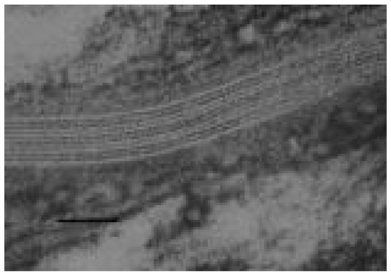
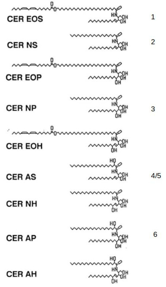
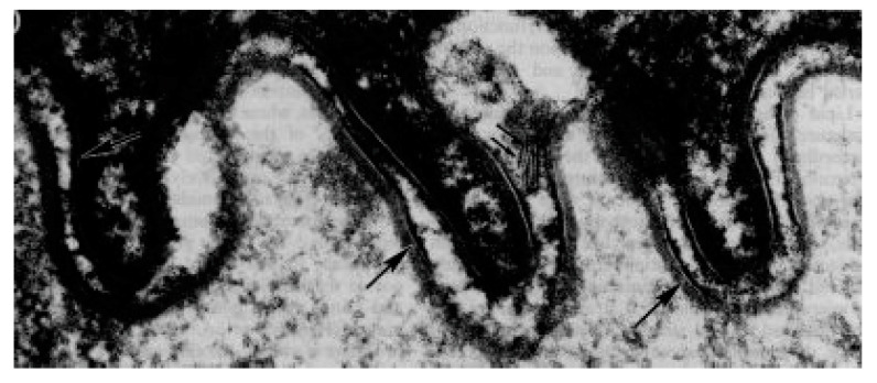
PubMed searches reveal much literature regarding lipids in barrier function of skin and less literature on lipids in barrier function of the oral mucosa. In terrestrial mammals, birds, and reptiles, the skin's permeability barrier is provided by ceramides, fatty acids, and cholesterol in the outermost layers of the epidermis, the stratum corneum. This layer consists of about 10-20 layers of cornified cells embedded in a lipid matrix. It effectively prevents loss of water and electrolytes from the underlying tissue, and it limits the penetration of potentially harmful substances from the environment. In the oral cavity, the regions of the gingiva and hard palate are covered by keratinized epithelia that much resemble the epidermis. The oral stratum corneum contains a lipid mixture similar to that in the epidermal stratum corneum but in lower amounts and is accordingly more permeable. The superficial regions of the nonkeratinized oral epithelia also provide a permeability barrier. These epithelial regions do contain ceramides, cholesterol, and free fatty acids, which may underlie barrier function. The oral epithelial permeability barriers primarily protect the underlying tissue by preventing the penetration of potentially toxic substances, including microbial products. Transdermal drug delivery, buccal absorption, and lipid-related disease are discussed.
Keywords: barrier function; ceramides; cholesterol; fatty acids; intercellular lamellae; keratinocytes; oral mucosa; skin.
Conflict of interest statement
The author has no conflict of interest.
Figures
References
-
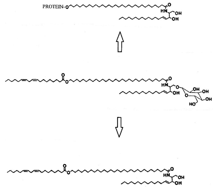
- Crumrine D., Khnykin D., Krieg P., Man M.-Q., Celli A., Mauro T.M., Wakefield J.S., Menon G., Mauldin E., Miner J.H., et al. Mutations in recessive congenital ichthoses illuminate the origin and functions of the corneocyte lipid envelope. J. Investig. Dermatol. 2019;139:760–768. doi: 10.1016/j.jid.2018.11.005. - DOI - PMC - PubMed
-
- Wertz P.W. Lipid metabolic events underlying the formation of the corneocyte lipid envelope. Skin Pharmacol. Physiol. 2021;34:38–50. - PubMed
-
- Attenborough D. Life on Earth. 1st ed. Little Brown & Company; Boston, MA, USA: 1980.
Publication types
MeSH terms
Substances
LinkOut - more resources
Full Text Sources
Medical
