. 2021 May 15;13(5):916.
doi: 10.3390/v13050916.

Persistent COVID-19 Symptoms Minimally Impact the Development of SARS-CoV-2-Specific T Cell Immunity

Affiliations

Persistent COVID-19 Symptoms Minimally Impact the Development of SARS-CoV-2-Specific T Cell Immunity

Hengsheng Fang et al. Viruses. .

Abstract

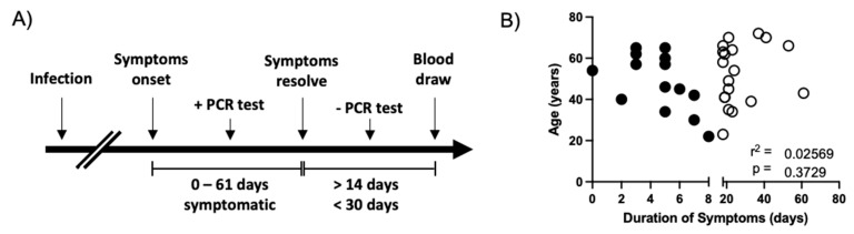
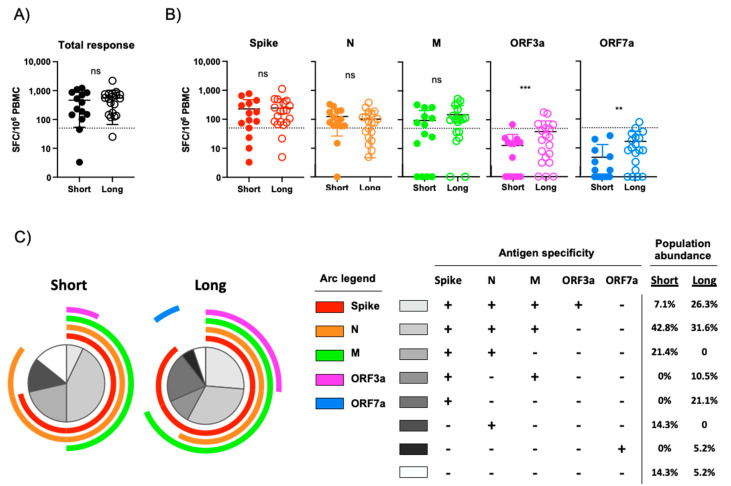
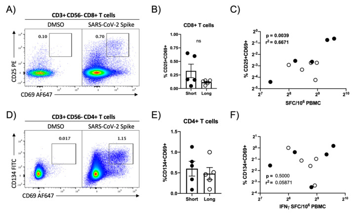
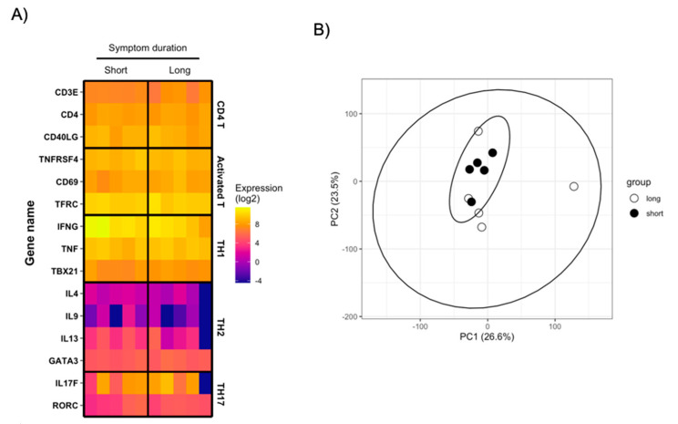
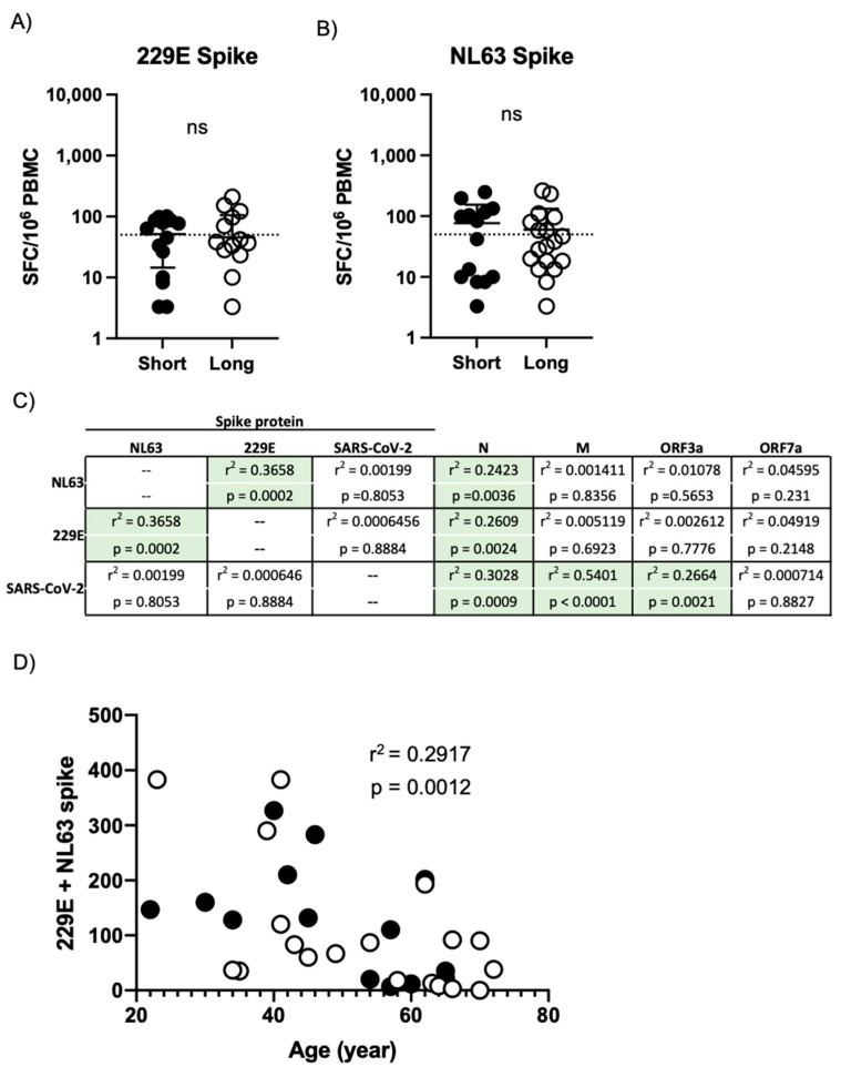
SARS-CoV-2 represents an unprecedented public health challenge. While the majority of SARS-CoV-2-infected individuals with mild-to-moderate COVID-19 resolve their infection with few complications, some individuals experience prolonged symptoms lasting for weeks after initial diagnosis. Persistent viral infections are commonly accompanied by immunologic dysregulation, but it is unclear if persistent COVID-19 impacts the development of virus-specific cellular immunity. To this end, we analyzed SARS-CoV-2-specific cellular immunity in convalescent COVID-19 patients who experienced eight days or fewer of COVID-19 symptoms or symptoms persisting for 18 days or more. We observed that persistent COVID-19 symptoms were not associated with the development of an overtly dysregulated cellular immune response. Furthermore, we observed that reactivity against the N protein from SARS-CoV-2 correlates with the amount of reactivity against the seasonal human coronaviruses 229E and NL63. These results provide insight into the processes that regulate the development of cellular immunity against SARS-CoV-2 and related human coronaviruses.

Keywords: COVID-19; SARS-CoV-2; T cells; cellular immunity; symptom duration.


Conflict of interest statement

S.J.T. reports other from Pfizer, during the conduct of the study; personal fees from Merck, personal fees from Sanofi, personal fees from Takeda, personal fees from Themisbio, personal fees from Janssen, outside the submitted work. All other authors declare that the research was conducted in the absence of any commercial or financial relationships that could be construed as a potential conflict of interest.

Figures

Figure 1
Figure 1
Study design and patient characteristics. (A) Schematic representation of the subject selection criteria used in this study and the timing of sample collection. (B) Analysis of the relationship between subject age and the duration of self-reported COVID-19 symptoms in all subjects included in this analysis. Asymptomatic SARS-CoV-2 PCR+ individuals were assigned 0 days of symptoms. Filled circles indicate subjects included in the short duration of symptoms group. Empty circles indicate individuals included in the long duration of symptoms group. r2 and p value calculated by 2-tailed Pearson Correlation test.
Figure 2
Figure 2
Assessment of SARS-CoV-2-specific cellular immunity by IFN-γ ELISPOT. (A) Total magnitude of SARS-CoV-2-specific cellular immunity in all study participants as defined by total reactivity against SARS-CoV-2 spike, N, M, ORF3a and ORF7a antigens. Subjects split by duration of self-reported symptoms. Dashed line indicates a 50 SFC/106 PBMC threshold for a positive response. (B) Magnitude of SARS-CoV-2-specific cellular immunity separated by major antigen in all study participants. Dashed line indicates a 50 SFC/106 PBMC threshold for a positive response. (C) Pattern of multi-antigen SARS-CoV-2 reactivity in all study subjects split by duration of self-reported symptoms. Arc color and arc length indicates reactivity against a given SARS-CoV-2 antigen. Internal plot wedge size indicates fraction of individuals with the indicated pattern of antigen reactivity. *** p < 0.001, ** p < 0.01 unpaired 2-tailed t test.
Figure 3
Figure 3
Flow cytometric quantification of SARS-CoV-2 spike protein-specific cellular immunity. (A) Representative flow cytometry plot demonstrating CD25 and CD69 upregulation in CD8+ T cells following in vitro SARS-CoV-2 spike protein peptide pool stimulation. (B) Magnitude of SARS-CoV-2 spike-specific CD8+ T cell responsiveness in select study participants split by duration of self-reported duration of COVID-19 symptoms. Plotted values are background subtracted from a total of 10 subjects. (C) Correlation analysis of SARS-CoV-2-specific cellular immunity as defined by CD8+ flow cytometry and IFN-γ ELISPOT. r2 and p value calculated by 2-tailed Pearson Correlation test. (D) Representative flow cytometry plot demonstrating CD134 and CD69 upregulation in CD4+ T cells following SARS-CoV-2 spike protein peptide pool stimulation. (E) Magnitude of SARS-CoV-2 spike-specific CD4+ T cell immunity in select study participants split by duration of self-reported duration of COVID-19 symptoms. Plotted values are background subtracted from a total of 10 subjects. (F) Correlation analysis of SARS-CoV-2-specific cellular immunity as defined by CD4+ flow cytometry and IFN-γ ELISPOT. r2 and p value calculated by 2-tailed Pearson correlation test.
Figure 4
Figure 4
Transcriptional analysis of SARS-CoV-2-reactive CD4+ T cells (A) Heatmap display of normalized gene expression in sorted SARS-CoV-2-reactive CD4+ T cells from total of 10 convalescent COVID-19 patients. Patients are separated by duration of self-reported COVID-19 symptoms. (B) PCA analysis of total normalized gene expression data from sorted SARS-CoV-2-reactive CD4+ T cells from total of 10 convalescent COVID-19 patients. Patients separated by duration of self-reported COVID-19 symptoms.
Figure 5
Figure 5
Assessment of seasonal human coronavirus cellular immunity in convalescent COVID-19 donors. (A) Magnitude of 229E spike protein-specific cellular immunity in all study participants split by duration of self-report COVID-19 symptoms. (B) Magnitude of NL63 spike protein-specific cellular immunity in all study participants split by duration of self-report COVID-19 symptoms. (C) Correlation table assessing the relationship between NL63, 229E, and SARS-CoV-2 spike protein reactivity in all subjects and the magnitude of reactivity against other human coronavirus antigens included in the study. r2 and p value calculated by 2-tailed Pearson Correlation test. (D) Relationship between subject age and total 229E/NL63 spike protein-specific cellular immune response. Filled circles indicate subjects included in the short duration of symptoms group. Empty circles indicate individuals included in the long duration of symptoms group. r2 and p value calculated by 2-tailed Pearson Correlation test.

References

    1. Lu R., Zhao X., Li J., Niu P., Yang B., Wu H., Wang W., Song H., Huang B., Zhu N., et al. Genomic characterisation and epidemiology of 2019 novel coronavirus: Implications for virus origins and receptor binding. Lancet. 2020;395:565–574. doi: 10.1016/S0140-6736(20)30251-8. - DOI - PMC - PubMed
    1. Zhu N., Zhang D., Wang W., Li X., Yang B., Song J., Zhao X., Huang B., Shi W., Lu R., et al. A Novel Coronavirus from Patients with Pneumonia in China, 2019. N. Engl. J. Med. 2020;382:727–733. doi: 10.1056/NEJMoa2001017. - DOI - PMC - PubMed
    1. Zhou P., Yang X.L., Wang X.G., Hu B., Zhang L., Zhang W., Si H.R., Zhu Y., Li B., Huang C.L., et al. A pneumonia outbreak associated with a new coronavirus of probable bat origin. Nature. 2020;579:270–273. doi: 10.1038/s41586-020-2012-7. - DOI - PMC - PubMed
    1. Guan W.J., Ni Z.Y., Hu Y., Liang W.H., Ou C.Q., He J.X., Liu L., Shan H., Lei C.L., Hui D.S.C., et al. Clinical Characteristics of Coronavirus Disease 2019 in China. N. Engl. J. Med. 2020;382:1708–1720. doi: 10.1056/NEJMoa2002032. - DOI - PMC - PubMed
    1. Huang C., Wang Y., Li X., Ren L., Zhao J., Hu Y., Zhang L., Fan G., Xu J., Gu X., et al. Clinical features of patients infected with 2019 novel coronavirus in Wuhan, China. Lancet. 2020;395:497–506. doi: 10.1016/S0140-6736(20)30183-5. - DOI - PMC - PubMed

Publication types

MeSH terms

Grants and funding