Review
. 2021 May 15;22(10):5240.
doi: 10.3390/ijms22105240.

Adenovirus Structure: What Is New?

Affiliations
Review

Adenovirus Structure: What Is New?

José Gallardo et al. Int J Mol Sci. .

Abstract

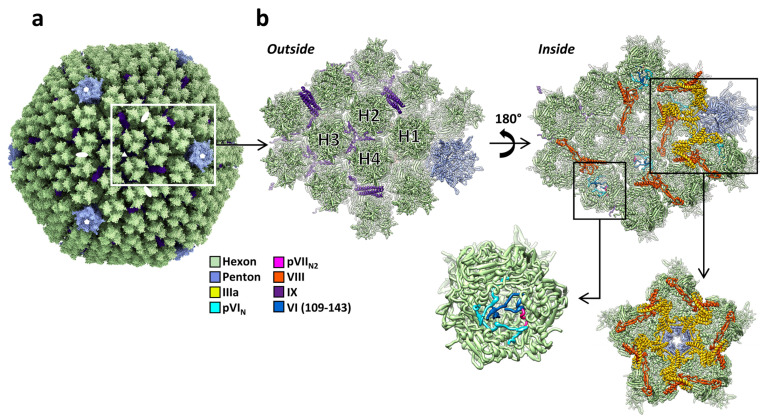
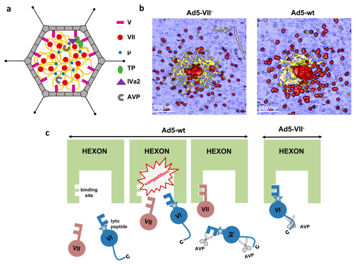
Adenoviruses are large (~950 Å) and complex non-enveloped, dsDNA icosahedral viruses. They have a pseudo-T = 25 triangulation number with at least 12 different proteins composing the virion. These include the major and minor capsid proteins, core proteins, maturation protease, terminal protein, and packaging machinery. Although adenoviruses have been studied for more than 60 years, deciphering their architecture has presented a challenge for structural biology techniques. An outstanding event was the first near-atomic resolution structure of human adenovirus type 5 (HAdV-C5), solved by cryo-electron microscopy (cryo-EM) in 2010. Discovery of new adenovirus types, together with methodological advances in structural biology techniques, in particular cryo-EM, has lately produced a considerable amount of new, high-resolution data on the organization of adenoviruses belonging to different species. In spite of these advances, the organization of the non-icosahedral core is still a great unknown. Nevertheless, alternative techniques such as atomic force microscopy (AFM) are providing interesting glimpses on the role of the core proteins in genome condensation and virion stability. Here we summarize the current knowledge on adenovirus structure, with an emphasis on high-resolution structures obtained since 2010.

Keywords: adenovirus; core proteins; cryo-EM; cryo-electron microscopy; crystallography; maturation; minor coat proteins; structure.


Conflict of interest statement

The authors declare no conflict of interest. The funders had no role in the design of the study; in the collection, analyses, or interpretation of data; in the writing of the manuscript; or in the decision to publish the results.

Figures

Figure 1
Figure 1
Structure of the AdV capsid. (a) General view of the HAdV-C5 capsid [28]. White symbols indicate the position of the icosahedral symmetry axes. The white rectangle highlights one icosahedral asymmetric unit. The fibers are not represented here, as they cannot be traced in studies using icosahedral symmetry. Structure rendering with ChimeraX [36]. (b) Zoom in on the asymmetric unit and its closest neighbors. Two views are provided: as seen from outside the capsid (left) and from inside (right). The four unique hexon trimers are labeled H1 to H4, and the different proteins are colored according to the color key at the left. One of the hexons is further magnified to show the pVIN and pVIIN2 peptides. Additionally highlighted is the GOS (penton plus peripentonal hexons), depicted as seen from inside the capsid. Structures rendered with Chimera [37].
Figure 2
Figure 2
Structure of the AdV capsid proteins. (a) Hexon trimer, with one of the monomers highlighted in vivid blue. (b) Penton base pentamer. The location of the untraced RGD loop in one of the monomers is indicated. In (a,b), the inner side of the particle would be at the bottom. (c) Localized reconstruction, without symmetry enforcement, of the HAdV-D26 fiber bound to the penton base. The view is from outside the capsid. An atomic model of the knob [19] is fitted into the density. Notice that the knob density appears clearly trimeric. The schematic diagram illustrates how the three-fold symmetric fiber (triangle) is shifted relative to the center (red dot) of the five-fold symmetric penton base (pentagon). Adapted from [47]. (d) Comparison between the LAdV-2 (yellow) and HAdV-C5 (gray) protein IIIa structures. Notice that the GOS-glue domains and part of the connecting helix overlap, but the VIII-binding domain in the LAdV-2 protein swings away from its position in the human virus. The black pentagon indicates the position of the 5-fold symmetry axis. Modified from [31]. (e) Schematics showing the organization of protein IX in HAdV-C5, BAdV-3, and HAdV-F41, and LH3 in LAdV-2. Molecules forming triskelions located at the center of the facet (I3 symmetry axis) are in cyan, and those located at the L3 axes in several shades of pink. The rope domains in HAdV-C5, and the rope and C-terminal domains in HAdV-F41, are depicted as dashed lines, indicating non-modeled residues. Monomers of protein IX/LH3 in the asymmetric unit are depicted on top of each schematic, with the N- and C-termini indicated. For HAdV-C5 and HAdV-F41, the four monomers in the AU are overlapped according to their triskelion region, to highlight the different conformations of the rope domain. Adapted from [30].
Figure 3
Figure 3
Roles of core protein VII in AdV assembly. (a) Schematic representation of the core with the dsDNA in yellow and proteins depicted according to the key at the right. (b) AFM images showing the core components released after breakage of Ad5-VII- and Ad5-wt particles. Material with a height consistent with dsDNA is yellow. Protein debris is in red. Reproduced from [91]. (c) Competition between proteins VI and VII for hexon binding impinges on AdV maturation and entry. Reproduced from [71].

References

    1. Rowe W.P., Huebner R.J., Gilmore L.K., Parrott R.H., Ward T.G. Isolation of a cytopathogenic agent from human adenoids undergoing spontaneous degeneration in tissue culture. Proc. Soc. Exp. Biol. Med. 1953;84:570–573. doi: 10.3181/00379727-84-20714. - DOI - PubMed
    1. Harrach B., Tarjan Z.L., Benko M. Adenoviruses across the animal kingdom: A walk in the zoo. FEBS Lett. 2019;593:3660–3673. doi: 10.1002/1873-3468.13687. - DOI - PubMed
    1. Berk A.J. Adenoviridae. In: Knipe D.M., Howley P.M., editors. Fields Virology. 6th ed. Volume 1. Wolters Kluwer/Lippincott Williams & Wilkins Health; Philadelphia, PA, USA: 2013. pp. 1704–1731.
    1. Kremer E.J. Pros and cons of adenovirus-based SARS-CoV-2 vaccines. Mol. Ther. J. Am. Soc. Gene Ther. 2020;28:2303–2304. doi: 10.1016/j.ymthe.2020.10.002. - DOI - PMC - PubMed
    1. Tumban E. Lead SARS-CoV-2 candidate vaccines: Expectations from phase III trials and recommendations post-vaccine approval. Viruses. 2020;13:54. doi: 10.3390/v13010054. - DOI - PMC - PubMed

LinkOut - more resources