. 2021 May 3;22(9):4841.
doi: 10.3390/ijms22094841.

Ribonucleic Acid Export 1 Is a Kinetochore-Associated Protein That Participates in Chromosome Alignment in Mouse Oocytes

Affiliations

Ribonucleic Acid Export 1 Is a Kinetochore-Associated Protein That Participates in Chromosome Alignment in Mouse Oocytes

Fan Chen et al. Int J Mol Sci. .

Abstract

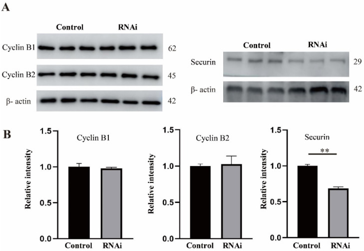
Ribonucleic acid export 1 (Rae1) is an important nucleoporin that participates in mRNA export during the interphase of higher eukaryotes and regulates the mitotic cell cycle. In this study, small RNA interference technology was used to knockdown Rae1, and immunofluorescence, immunoblotting, and chromosome spreading were used to study the role of Rae1 in mouse oocyte meiotic maturation. We found that Rae1 is a crucial regulator of meiotic maturation of mouse oocytes. After the resumption of meiosis (GVBD), Rae1 was concentrated on the kinetochore structure. The knockdown of Rae1 by a specific siRNA inhibited GVBD progression at 2 h, finally leading to a decreased 14 h polar body extrusion (PBE) rate. However, a comparable 14 h PBE rate was found in the control, and the Rae1 knockdown groups that had already undergone GVBD. Furthermore, we found elevated PBE after 9.5 h in the Rae1 knockdown oocytes. Further analysis revealed that Rae1 depletion significantly decreased the protein level of securin. In addition, we detected weakened kinetochore-microtubule (K-MT) attachments, misaligned chromosomes, and an increased incidence of aneuploidy in the Rae1 knockdown oocytes. Collectively, we propose that Rae1 modulates securin protein levels, which contribute to chromosome alignment, K-MT attachments, and aneuploidy in meiosis.

Keywords: Rae1; aneuploidy; chromosome alignment; meiotic maturation; mouse oocyte; securin.


Conflict of interest statement

The authors declare that no competing interests exist.

Figures

Figure 1
Figure 1
The subcellular localization and protein expression pattern of Rae1 during oocyte meiotic maturation. (A) Representative images of the subcellular localization of Rae1 in oocyte at the germinal vesicle (GV), metaphase I (MI), and metaphase II (MII) stages. Rae1 is shown in red, α-tubulin in green, and 4’,6-diamidino-2-phenylindole (DAPI) in blue. Scale bar = 10 μm. (B) Representative images of Rae1 localization in chromosomes of oocytes at the germinal vesicle breakdown (GVBD), MI, and MII stage by chromosome spreading. Rae1 is shown in red and DAPI in blue. Scale bar = 5 μm. (C). The co-localization of Rae1 and CREST in chromosomes of the MIIstage oocyte by chromosome spreading. Rae1 is shown in red, CREST in green, and DAPI in blue. Scale bar = 5 μm. (D) The protein level of Rae1 at the GV, GVBD, MI, and MII stages detected by Western blotting. (E) Relative intensity of Rae1 at the GV, GVBD, MI, and MII stages is presented, which were calculated by Rae1/tubulin. NS means no significant difference. ** p < 0.01. The data are presented as mean ± standard error of the mean (SEM) of three independent experiments.
Figure 2
Figure 2
Rae1 knockdown affects the meiotic maturation process. (A) Efficiency of Rae1–RNAi after siRNA microinjection was verified by immunoblotting. (B) RNAi after siRNA microinjection was verified by immunostaining. Rae1 is shown in red and DAPI in blue. Scale bar = 20 μm. (C) The GVBD rates in the control group and Rae1 knockdown group were recorded 2 h after release from 3-Isobutyl-1-Methylxanthine (IBMX) arrest. * p < 0.05. (D). The PBE rates in the control group and Rae1 knockdown group were recorded 14 h after release from IBMX arrest. * p < 0.05. (E) The PBE rates in the control group and Rae1 knockdown group were recorded after 14 h in 2 h GVBD-resumed oocytes after release from IBMX arrest. p > 0.05. (F) The PBE rates in the control group and Rae1 knockdown group were recorded after 9 h in 2 h GVBD-resumed oocytes after release from IBMX arrest. * p < 0.05. The data are presented as mean ± SEM of at least three independent experiments.
Figure 3
Figure 3
Rae1 knockdown induces abnormal chromosome alignment and elevated aneuploid rate in oocytes. (A) Representative confocal images of spindle morphology and chromosome alignment in the control and Rae1 knockdown MI oocytes. The white line and arrow indicate that in the control group, chromosomes are congressed and aligned at the spindle midzone, while in the Rae1-knockdown groups, chromosomes are scattered and dispersed. α-tubulin is shown in green and DAPI is shown in blue. Scale bar = 10 μm. (B) The rate of misaligned chromosomes is recorded and compared in the control (n = 96) and Rae1 knockdown MI oocytes (n = 108). ** p < 0.01 (C) Representative confocal image of chromosome spread at the MII stage in the control and Rae1 knockdown group. Control oocytes with a normal haploid complement of 20 chromosomes and Rae1 knockdown oocyte with 21 chromosomes. Chromosomes were stained with propidium iodide (PI) and is shown in red. Scale bar = 5 μm. (D) The aneuploidy rate is recorded and compared in the control (n = 31) and Rae1 knockdown oocytes (n = 35). ** p < 0.01. The data are presented as mean ± SEM of at least three independent experiments.
Figure 4
Figure 4
Rae1 knockdown weakens kinetochore–microtubule (K-MT) attachment in oocytes. (A) Representative confocal image of K-MT attachment in the control and Rae1 knockdown MI oocytes after cold treatment. α-tubulin is shown in green, CREST in red, and DAPI in blue. White arrows indicate nonconnected kinetochores in the Rae1 knockdown oocytes. Scale bar = 5 μm. (B) The rate of unattached kinetochores was recorded and compared in the control (n = 125) and Rae1 knockdown oocytes (n = 143). ** p < 0.01. The data are presented as mean ± SEM of at least three independent experiments.
Figure 5
Figure 5
The location of BubR1 and Mad1 was not affected in the Rae1 knockdown oocytes. After Rae1 knockdown, GV stage oocytes were cultured for 8.5 h and then collected for immunofluorescent staining of BubR1 and Mad1. (A) Confocal image of BubR1 localization in the control and Rae1 knockdown oocytes by chromosome spreading. BubR1 is shown in green and DAPI in blue. Scale Bar = 5 μm. (B) Confocal image of Mad1 localization in the control and Rae1 knockdown oocytes by chromosome spreading. Mad1 is shown in red and DAPI in blue. Scale Bar = 5 μm.
Figure 6
Figure 6
Rae1 knockdown significantly decreases the protein level of securin in MI stage oocytes. After Rae1 knockdown, GV stage oocytes were cultured for 8 h (MI stage) after release from IBMX arrest and were collected for the Western blotting of cyclin B1, cyclin B2, and securin. (A) Three independent repeat results of securin, cyclin B1, and cyclin B2 by Western blotting. (B) The relative intensity of securin, cyclin B1, and cyclin B2 were compared in the control group and Rae1 knockdown group. ** p < 0.01. The data are presented as mean ± SEM of at least three independent experiments.

Similar articles

Cited by

References

    1. Cronshaw J.M., Krutchinsky A.N., Zhang W., Chait B.T., Matunis M.J. Proteomic analysis of the mammalian nuclear pore complex. J. Cell Biol. 2002;158:915–927. doi: 10.1083/jcb.200206106. - DOI - PMC - PubMed
    1. Brown J.A., Bharathi A., Ghosh A., Whalen W., Fitzgerald E., Dhar R. A mutation in the Schizosaccharomyces pombe rae1 gene causes defects in poly(A)+ RNA export and in the cytoskeleton. J. Biol. Chem. 1995;270:7411–7419. doi: 10.1074/jbc.270.13.7411. - DOI - PubMed
    1. Murphy R., Watkins J.L., Wente S.R. GLE2. A Saccharomyces cerevisiae homologue of the Schizosaccharomyces pombe export factor RAE1, is required for nuclear pore complex structure and function. Mol. Biol. Cell. 1996;7:1921–1937. doi: 10.1091/mbc.7.12.1921. - DOI - PMC - PubMed
    1. Whalen W.A., Bharathi A., Danielewicz D., Dhar R. Advancement through mitosis requires rae1 gene function in fission yeast. Yeast. 1997;13:1167–1179. doi: 10.1002/(SICI)1097-0061(19970930)13:12<1167::AID-YEA154>3.0.CO;2-O. - DOI - PubMed
    1. Blower M.D., Nachury M., Heald R., Weis K.A. Rae1-containing ribonucleoprotein complex is required for mitotic spindle assembly. Cell. 2005;121:223–234. doi: 10.1016/j.cell.2005.02.016. - DOI - PubMed

MeSH terms

Substances

LinkOut - more resources