

Parametric Imaging of Contrast-Enhanced Ultrasound (CEUS) for the Evaluation of Acute Gastrointestinal Graft-Versus-Host Disease
- PMID: 34063640
- PMCID: PMC8147630
- DOI: 10.3390/cells10051092
Parametric Imaging of Contrast-Enhanced Ultrasound (CEUS) for the Evaluation of Acute Gastrointestinal Graft-Versus-Host Disease
Abstract
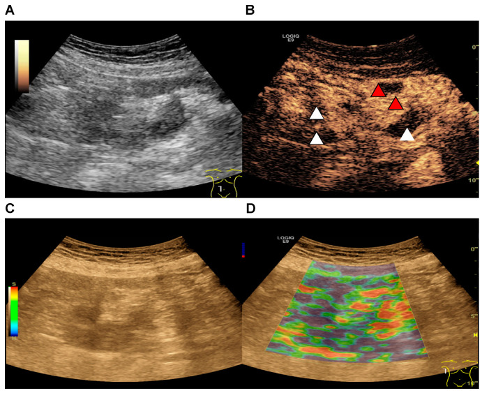
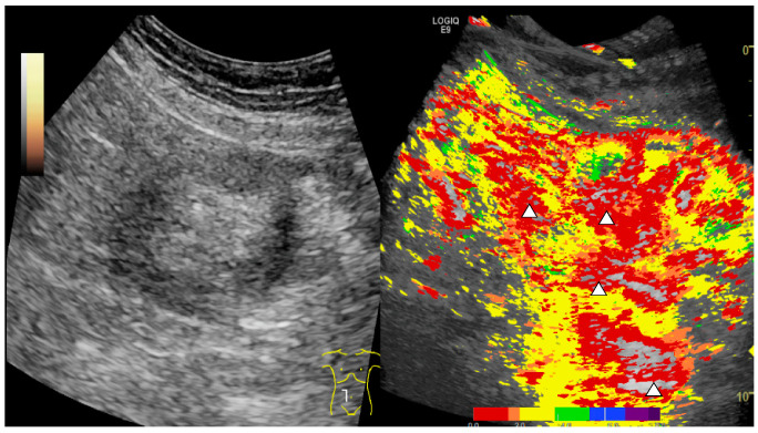
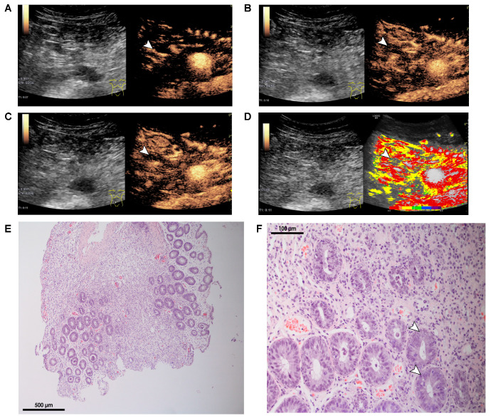
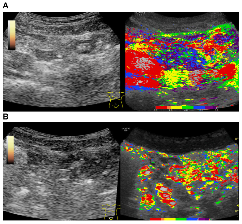
In recent years contrast-enhanced ultrasound (CEUS) has been an emerging diagnostic modality for the detection of acute gastrointestinal (GI) graft-versus-host disease (GvHD) in patients after allogeneic stem cell transplantation. However, broad clinical usage has been partially limited by its high dependence on the expertise of an experienced examiner. Thus, the aim of this study was to facilitate detection of acute GI GvHD by implementing false color-coded parametric imaging of CEUS. As such, two inexperienced examiners with basic knowledge in abdominal and vascular ultrasound analyzed parametric images obtained from patients with clinical suspicion for acute GvHD in a blinded fashion. As diagnostic gold standard, histopathological GvHD severity score on intestinal biopsies obtained from lower GI tract endoscopy was performed. The evaluation of parametric images by the two inexperienced ultrasound examiners in patients with histological confirmation of acute GI GvHD was successful in 17 out of 19 patients (89%) as opposed to analysis of combined B-mode ultrasound, strain elastography, and CEUS by an experienced examiner, which was successful in 18 out of 19 of the patients (95%). Therefore, CEUS with parametric imaging of the intestine was technically feasible and has the potential to become a valuable diagnostic tool for rapid and widely accessible detection of acute GvHD in clinical practice.
Keywords: CEUS; GvHD; allogeneic stem cell transplantation; dynamic vascularization; parametric imaging.
Conflict of interest statement
The authors declare no conflict of interest.
Figures
Similar articles
-
Is Contrast-Enhanced Ultrasonography a New, Reliable Tool for Early-Graft-versus-Host Disease Diagnosis?J Clin Med. 2024 Oct 11;13(20):6065. doi: 10.3390/jcm13206065. J Clin Med. 2024. PMID: 39458018 Free PMC article. Review.
-
Non-invasive diagnosis of acute intestinal graft-versus-host disease by a new scoring system using ultrasound morphology, compound elastography, and contrast-enhanced ultrasound.Bone Marrow Transplant. 2019 Jul;54(7):1038-1048. doi: 10.1038/s41409-018-0381-4. Epub 2018 Nov 6. Bone Marrow Transplant. 2019. PMID: 30401964 Clinical Trial.
-
Contrast-enhanced ultrasound for differential diagnosis of suspected GvHD in patients after allogeneic transplantation.Clin Hemorheol Microcirc. 2011;49(1-4):129-36. doi: 10.3233/CH-2011-1463. Clin Hemorheol Microcirc. 2011. PMID: 22214684 Clinical Trial.
-
Magnetic resonance enterography for assessment of intestinal graft-versus-host disease after allogeneic stem cell transplantation.Eur Radiol. 2015 May;25(5):1229-37. doi: 10.1007/s00330-014-3503-4. Epub 2014 Nov 23. Eur Radiol. 2015. PMID: 25417129
-
New insight for the diagnosis of gastrointestinal acute graft-versus-host disease.Mediators Inflamm. 2014;2014:701013. doi: 10.1155/2014/701013. Epub 2014 Mar 11. Mediators Inflamm. 2014. PMID: 24733964 Free PMC article. Review.
Cited by
-
Super-Resolution Contrast-Enhanced Ultrasound Examination Down to the Microvasculature Enables Quantitative Analysis of Liver Lesions: First Results.Life (Basel). 2025 Jun 20;15(7):991. doi: 10.3390/life15070991. Life (Basel). 2025. PMID: 40724493 Free PMC article.
-
Advances in the clinical diagnosis of lung cancer using contrast-enhanced ultrasound.Front Med (Lausanne). 2025 Mar 19;12:1543033. doi: 10.3389/fmed.2025.1543033. eCollection 2025. Front Med (Lausanne). 2025. PMID: 40177283 Free PMC article. Review.
-
Is Contrast-Enhanced Ultrasonography a New, Reliable Tool for Early-Graft-versus-Host Disease Diagnosis?J Clin Med. 2024 Oct 11;13(20):6065. doi: 10.3390/jcm13206065. J Clin Med. 2024. PMID: 39458018 Free PMC article. Review.
-
Current Developments and Role of Intestinal Ultrasound including the Advent of AI.Diagnostics (Basel). 2024 Apr 3;14(7):759. doi: 10.3390/diagnostics14070759. Diagnostics (Basel). 2024. PMID: 38611672 Free PMC article. Review.
-
Enhanced Diagnostic Imaging: Arrival-Time Parametric Imaging in Contrast-Enhanced Ultrasound for Multi-Organ Assessment.Med Sci Monit. 2024 Nov 28;30:e945281. doi: 10.12659/MSM.945281. Med Sci Monit. 2024. PMID: 39604210 Free PMC article. Review.
References
Publication types
MeSH terms
Substances
Grants and funding
LinkOut - more resources
Full Text Sources
Medical
Research Materials
