Review
. 2021 May 21;22(11):5412.
doi: 10.3390/ijms22115412.

α-Helices in the Type III Secretion Effectors: A Prevalent Feature with Versatile Roles

Affiliations
Review

α-Helices in the Type III Secretion Effectors: A Prevalent Feature with Versatile Roles

Anastasia D Gazi et al. Int J Mol Sci. .

Abstract

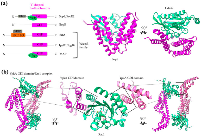
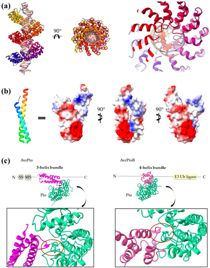
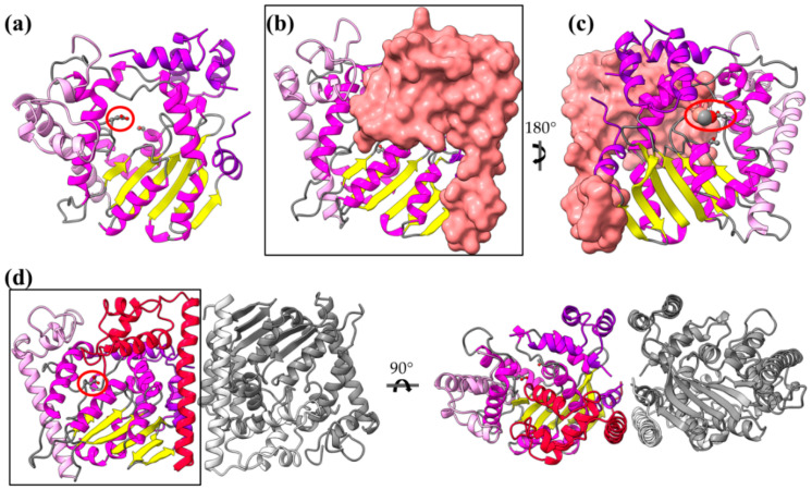
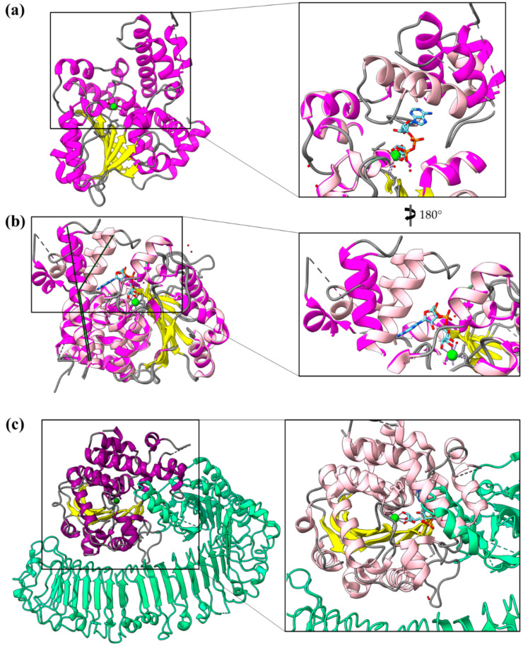
Type III Secretion Systems (T3SSs) are multicomponent nanomachines located at the cell envelope of Gram-negative bacteria. Their main function is to transport bacterial proteins either extracellularly or directly into the eukaryotic host cell cytoplasm. Type III Secretion effectors (T3SEs), latest to be secreted T3S substrates, are destined to act at the eukaryotic host cell cytoplasm and occasionally at the nucleus, hijacking cellular processes through mimicking eukaryotic proteins. A broad range of functions is attributed to T3SEs, ranging from the manipulation of the host cell's metabolism for the benefit of the bacterium to bypassing the host's defense mechanisms. To perform this broad range of manipulations, T3SEs have evolved numerous novel folds that are compatible with some basic requirements: they should be able to easily unfold, pass through the narrow T3SS channel, and refold to an active form when on the other side. In this review, the various folds of T3SEs are presented with the emphasis placed on the functional and structural importance of α-helices and helical domains.

Keywords: 4-α-helix bundle; Leucine-Rich Repeat (LRR); Novel E3 Ligase (NEL); Transcription Activator-Like Effector (TALE); Type III Secretion System (T3SS); Type III Secretion effector (T3SE); coiled coil; dictionary of secondary structure in proteins (DSSP).


Conflict of interest statement

The authors declare no conflict of interest.

Figures

Figure 1
Figure 1
Overview of a T3S nanomachine embedded in the two bacterial membranes. Early and middle secretion substrates are almost all α-helical. The early secretion substrates build the inner rod adaptor (yellow α-helices just above the secretion core, PDB id 6RWY) and the needle (in orange, EMDB id 2803, red α-helices, PDB id 6RWY) or pilus of the nanomachinery that extends to the extracellular space. In animal pathogenic bacteria, the needle is capped with the pentameric needle tip (here in blue, EMDB id 2803), a middle stage substrate. Enteropathogenic (EPEC)/enterohemorrhagic (EHEC) Escherichia coli produce thicker extracellular appendages termed filaments (here in purple, PDB id 7KHW). The rest of the parts of the T3S nanomachine, that are not formed from secretion substrates, are depicted in shades of grey to white (PDB id: 6Q15, EMDB ids: 20561, 20611). In the left column, high resolution structures in cartoon representation (α-helices in magenta, β-strands in yellow) corresponding to early secretion substrates that polymerize to build the corresponding T3S parts shown in the right column. Chaperones to maintain early secretion substrates inside the bacterial cytoplasm have also been described (here represented in different shades of grey: light grey for chaperones with tetratrico-peptide repeats (TPR), grey for the rest). Translocators are also considered middle stage secretion substrates as they must be secreted before the effectors. The major subunit of the translocation pore is maintained inside the bacterial cytoplasm in a secretion competent folding state. The chaperone of the translocator, which also possesses TPR, anchors to the N-terminal Chaperone Binding Domain (CBD) of the translocator, while also covering the transmembrane helices of the translocator by extensively interacting with it [13]. IpaBcc denotes the known long coiled coil domain of IpaB. The CBD is proceeding this domain, while the transmembrane helices are following this domain and shown here protected by the chaperone in an analogy to the AcrH/AopB case [14]. PDB ids used for the left column: 3WXX, 5WKQ, 6RWY, 2IZP, 2P58, 1XOU.
Figure 2
Figure 2
A T3S effector trapped inside the secretion tunnel. The T3SS needle complex atomic structure of Salmonella enterica was determined in two functional states. Here with the secretion substrate trapped inside (PDB id 7ahi) [21]. (a) A calculated 5 Å map of the atomic model is shown for simplicity. Substrate is displayed in magenta, T3SS needle complex in light purple. (b) Zoom on the needle and the secretion tunnel. (c,d) Smoothed surface of the tunnel displayed in light blue. The tunnel has a right-handed helical grooved surface and is wide enough to allow secretion substrates to form α-helices. In (c) the molecular surface representation of the substrate is shown. In (d) the cartoon representation is shown.
Figure 3
Figure 3
Representative structures for T3SEs. T3SEs are shown in cartoon representation with α-helices in magenta, β-strands in yellow, rest of secondary structure in grey. N-terminal and C-terminal unstructured parts are depicted here in grey broken lines. Dimeric T3SS class I chaperones (or chaperones of effectors) are displayed in grey cartoon representation. Host protein targets that have been co-crystallized with the T3SEs are shown in surface representation in green color. In blue, the alternative conformation of YopH Chaperone Binding Domain (CBD), in the absence of the cognate chaperone. PDB ids used: 1JL5, 4PUF, 4O96, 4O2I, 2QKW, 2NUD, 4FMB, 1GZS, 5CPC, 1S21, 3TU3, 6ACI, 1HE1, 1XXP, 6GNN, 7JLU, 2YPF, 6HQZ, 4RSW, 5T09, 6PWD.
Figure 4
Figure 4
The secondary structure composition of T3SE domains in comparison to PDB. The average occurrence of α-helices in T3SEs is approximately 10% higher compared to the PDB proteome [25]. However, the information from the T3SE-determined crystal structures usually comes from the structured domains of the T3SEs, while their frequently unstructured/flexible N-terminal and C-terminal parts are usually missing from the determined crystal structures. Secondary structure assignments were performed for each PDB id presented in Figure 3.
Figure 5
Figure 5
CopN gatekeeper structure. (a) The CopN structure consisting of three (R1–R3) structurally homologous 5-helix assemblies, shown in different magenta shades. (b) Close-up of the R2 and R3 motifs of CopN. The view of panel (b) is rotated 90 degrees in comparison with the view in panel (a). The pair of parallel helices is shown in yellow for clarity. (c) and (d) Part of the CopN structure (only the R3 and R2 motifs are shown) in complex with the Scc3 chaperone of translocators (cyan) and the tubulin (green), respectively.
Figure 6
Figure 6
ExoU structure (multi-colored) in complex with the SpcU chaperone (gray). The complex is shown in two views 180 degrees apart. At the right panel the molecule has been clipped for the emphasis to be given on the 4-helix-bundle (magenta). ExoU comprises a highly unstructured N-terminal domain (red), which interacts with the chaperone, a catalytic, patatin-like domain (green), an all-helical bridging domain (blue) and a C-terminal 4-helix domain (magenta). The 4-α-helix-bundle domain is involved in the membrane and ubiquitin binding. The corresponding binding sites are indicated. Ub stands for ubiquitin.
Figure 7
Figure 7
Bacterial structures with GAP activities. (a) Schematic comparison of domain architecture of YopE, ExoS/T and SptP. (b) The GAP domain is a 4-helix-bundle shown here in the SptP/Rac1 complex structure. Rac1 is shown in orange, the GAP domain in pink and the extra catalytic activity of tyrosine phosphatase (PTPase) in green. (c) Close-up of the interaction interface between the Rac1 and GAP domains. MLD stands for membrane localization domain, SS for secretion sequence, CB for chaperone binding.
Figure 8
Figure 8
Bacterial structures with GAP activities. (a) Schematic representation of the domain architecture of EspG/VirA family members. (b) The homodimer of VirA. One monomer is colored white and the other with three different colors highlighting the individual domains. The close-up emphasizes the dimer interface. (c) In the EspG/Rab1 complex, the EspG color code is similar to the one used for VirA, and Rab 1 is colored orange. The close-up emphasizes the complex interaction interface and shows that Rab1 binds on a site equivalent to those used for dimerization. MLD stands for membrane localization domain, SS for secretion sequence, CB for chaperone binding.
Figure 9
Figure 9
Bacterial structures with GEF (a) and GDI (b) activities. (a) Left: Schematic comparison of the domain architecture of bacterial GEFs. SKIP stands for the host SifA kinesin interacting protein. Right: The GEF domain is a V-shaped helical bundle as it is representatively illustrated by the SopE structure (magenta). The structure of SopE is shown in complex with the host Cdc42 (green, PDB id 1GZS). (b)The YopO/YpkA dimer structure (magenta/pink) in complex with Rac1 (green, PDB id 2H7V).
Figure 10
Figure 10
(a) PthXo1 protein in complex with DNA (PDB id 3UGM). The structure comprises tandem repeats of a helix–loop–helix motif. The protein is colored from N-terminus (blue) to C-terminus (red). DNA specific recognition and binding occurs through the hypervariable, in sequence, loop of the motif (right). (b) The AvrRps4 mature structure is a coiled coil with electrostatically diverse sides (PDB id 4B6X). Red and blue colors on the surface denote negative and positive charge, respectively. (c) Each of the AvrPto and AvrPtoB proteins (magenta) interact with the Pto host kinase (green). Left: schematic diagram of the AvrPto protein and close-up of the AvrPto–Pto interaction (PDB id 2QKW). Right: schematic diagram of the AvrPtoB protein and close-up of the AvrPtoB–Pto interaction (PDB id 3HGK).
Figure 11
Figure 11
The structure of AvrRxo1-ORF1 and its complex with the AvrRxo1-ORF2 chaperone (PDB id 4Z8V). (a) The AvrRxo1-ORF1 monomer consists of a major, middle α/β domain (helices are colored in magenta) and two flanking domains, whose helices are colored pink and violet, respectively. (b,c) The 1:1 complex of AvrRxo1-ORF1/AvrRxo1-ORF2 in two views, 180 degrees apart. AvrRxo1-ORF2 is shown in surface representation. (d) The 2:2 complex of AvrRxo1-ORF1/AvrRxo1-ORF2 in two views, 90 degrees apart. One dimer is gray and the other is colorful, consistent with colors in (a). AvrRxo1-ORF2 is shown in red cartoon.
Figure 12
Figure 12
XopQ structure and conformational changes. (a) The structure of Xanthomonas XopQ is a Rossmann fold implemented with a mobile helical segment able to function as a lid of the active site upon substrate binding (PDB id 4KL0). (b) Closure of the active site is achieved via a bend which occurs in the middle of a long helix (PDB id 4P5F). The dark and light tones of magenta indicate the open and closed conformations of XopQ protein, respectively. (c) Left panel: The structure of XopQ (magenta/yellow) in complex with the Roq1 (green) indicates that the latter inserts a helical segment into the active site cleft of XopQ (PDB id 7JLU). Right panel: Close-up of the complex. In the structure, the XopQ has been substituted by a superimposed closed conformation. It is evident that the Roq1 helix insertion overlaps with the XopQ helical-lid in the closed conformation.

References

    1. Portaliou A.G., Tsolis K.C., Loos M.S., Zorzini V., Economou A. Type III Secretion: Building and Operating a Remarkable Nanomachine. Trends Biochem. Sci. 2016;41:175–189. doi: 10.1016/j.tibs.2015.09.005. - DOI - PubMed
    1. Tampakaki A.P., Skandalis N., Gazi A.D., Bastaki M.N., Sarris P.F., Charova S.N., Kokkinidis M., Panopoulos N.J. Playing the “Harp”: Evolution of our understanding of hrp/hrc genes. Annu. Rev. Phytopathol. 2010;48:347–370. doi: 10.1146/annurev-phyto-073009-114407. - DOI - PubMed
    1. Abby S.S., Rocha E.P.C. The Non-Flagellar Type III Secretion System Evolved from the Bacterial Flagellum and Diversified into Host-Cell Adapted Systems. PLoS Genet. 2012;8:e1002983. doi: 10.1371/journal.pgen.1002983. - DOI - PMC - PubMed
    1. Lunelli M., Kamprad A., Bürger J., Mielke T., Spahn C.M.T., Kolbe M. Cryo-EM structure of the Shigella type III needle complex. PLoS Pathog. 2020;16:e1008263. doi: 10.1371/journal.ppat.1008263. - DOI - PMC - PubMed
    1. Hu B., Morado D.R., Margolin W., Rohde J.R., Arizmendi O., Picking W.L., Picking W.D., Liu J. Visualization of the type III secretion sorting platform of Shigella flexneri. Proc. Natl. Acad. Sci. USA. 2015;112:1047–1052. doi: 10.1073/pnas.1411610112. - DOI - PMC - PubMed

MeSH terms

Substances

LinkOut - more resources