

Microglial Extracellular Vesicles as Vehicles for Neurodegeneration Spreading
- PMID: 34063832
- PMCID: PMC8224033
- DOI: 10.3390/biom11060770
Microglial Extracellular Vesicles as Vehicles for Neurodegeneration Spreading
Abstract
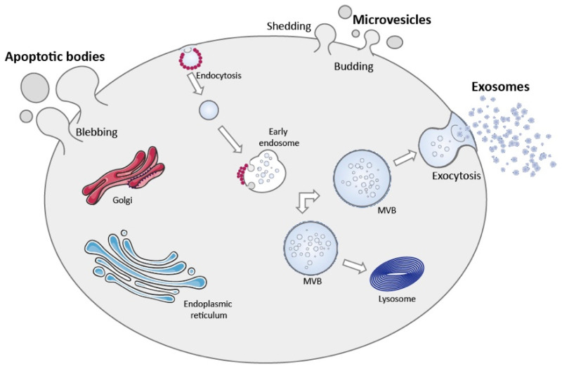
Microglial cells are the neuroimmune competent cells of the central nervous system. In the adult, microglia are responsible for screening the neuronal parenchyma searching for alterations in homeostasis. Chronic neuroinflammation plays a role in neurodegenerative disease. Indeed, microglia-mediated neuroinflammation is involved in the onset and progression of several disorders in the brain and retina. Microglial cell reactivity occurs in an orchestrated manner and propagates across the neural parenchyma spreading the neuroinflammatory signal from cell to cell. Extracellular vesicles are important vehicles of intercellular communication and act as message carriers across boundaries. Extracellular vesicles can be subdivided in several categories according to their cellular origin (apoptotic bodies, microvesicles and exosomes), each presenting, different but sometimes overlapping functions in cell communication. Mounting evidence suggests a role for extracellular vesicles in regulating microglial cell action. Herein, we explore the role of microglial extracellular vesicles as vehicles for cell communication and the mechanisms that trigger their release. In this review we covered the role of microglial extracellular vesicles, focusing on apoptotic bodies, microvesicles and exosomes, in the context of neurodegeneration and the impact of these vesicles derived from other cells in microglial cell reactivity.
Keywords: exosomes; extracellular vesicles; microglia; microvesicles; neurodegeneration.
Conflict of interest statement
The authors declare no conflict of interest.
Figures
References
Publication types
MeSH terms
LinkOut - more resources
Full Text Sources
Medical
