Review
. 2021 May 21;10(11):2235.
doi: 10.3390/jcm10112235.

Myasthenia Gravis: Epidemiology, Pathophysiology and Clinical Manifestations

Affiliations
Review

Myasthenia Gravis: Epidemiology, Pathophysiology and Clinical Manifestations

Laura Dresser et al. J Clin Med. .

Abstract

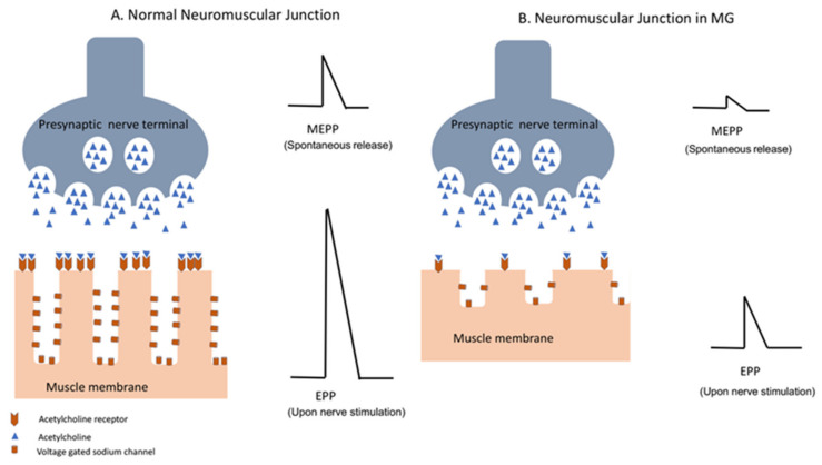
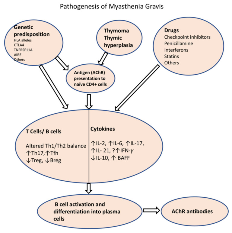
Myasthenia gravis (MG) is an autoimmune neurological disorder characterized by defective transmission at the neuromuscular junction. The incidence of the disease is 4.1 to 30 cases per million person-years, and the prevalence rate ranges from 150 to 200 cases per million. MG is considered a classic example of antibody-mediated autoimmune disease. Most patients with MG have autoantibodies against the acetylcholine receptors (AChRs). Less commonly identified autoantibodies include those targeted to muscle-specific kinase (MuSK), low-density lipoprotein receptor-related protein 4 (Lrp4), and agrin. These autoantibodies disrupt cholinergic transmission between nerve terminals and muscle fibers by causing downregulation, destruction, functional blocking of AChRs, or disrupting the clustering of AChRs in the postsynaptic membrane. The core clinical manifestation of MG is fatigable muscle weakness, which may affect ocular, bulbar, respiratory and limb muscles. Clinical manifestations vary according to the type of autoantibody, and whether a thymoma is present.

Keywords: B cells; T cells; acetylcholine receptor; autoantibodies; cytokines; myasthenia gravis.


Conflict of interest statement

K.R. has received honoraria for consultations and serving on advisory boards for Alexion, Kabafusion and Grifols. B.S. and K.R. have received funding from Alexion for conducting clinical trials on MG and ALS. L.D. and R.W. declare no conflict of interest.

Figures

Figure 1
Figure 1
Diagram depicting the secretion of agrin from the presynaptic membrane and its interaction with Lrp4, which results in reorganization and reorientation of MuSK, promoting a signaling pathway that leads to synaptic differentiation, including clustering of AChRs. This involves recruiting rapsyn which links AChRs to the cytoskeleton (not shown).
Figure 2
Figure 2
Neuromuscular transmission in normal individuals (A) and in patients with MG (B). Decreased density of the AChR and complement-mediated damage to the postsynaptic membrane in MG patients result in decrease in miniature end plate potential (MEPP), which occurs with spontaneous release of AChR vesicles, as well as endplate potential (EPP) in response to nerve action potential of the presynaptic membrane. Diminished amplitude of EPP in MG results in impaired neuromuscular transmission.
Figure 3
Figure 3
Schematic diagram of pathogenesis of AChR-MG. Impaired tolerance to the AChR is the result of thymoma, thymic dysplasia or due to certain genetic background, which results in presentation of AChR to the naïve T cells by thymic myoid cells or antigen-presenting cells. Among the environmental factors, certain drugs are known to cause de novo MG through alterations of immune homeostasis (drug-induced MG is extensively covered in another paper in this special edition). A number of T cell and B cell subtypes and their cytokines play roles in perturbation of immune homeostasis that results in production of ACR antibodies. HLA: Human Leucocyte antigen; CTLA4: cytotoxic T-lymphocyte-associated protein 4; TNFRSF11A: tumor necrosis factor receptor 4 superfamily, member 11a; AIRE: autoimmune regulator; Th1: T helper 1; Th2: T helper 2; Tfh: T follicular helper; Treg: regulatory T cell; Bregs: regulatory B cells; IL: interleukin; BAFF: B cell activating factor.

References

    1. McGrogan A., Sneddon S., de Vries C.S. The Incidence of Myasthenia Gravis: A Systematic Literature Review. Neuroepidemiology. 2010;34:171–183. doi: 10.1159/000279334. - DOI - PubMed
    1. Patrick J., Lindstrom J. Autoimmune Response to Acetylcholine Receptor. Science. 1973;180:871–872. doi: 10.1126/science.180.4088.871. - DOI - PubMed
    1. Fambrough D.M., Drachman D.B., Satyamurti S. Neuromuscular Junction in Myasthenia Gravis: Decreased Acetylcholine Receptors. Science. 1973;182:293–295. doi: 10.1126/science.182.4109.293. - DOI - PubMed
    1. McConville J., Farrugia M.E., Beeson D., Kishore U., Metcalfe R., Newsom-Davis J., Vincent A. Detection and Characterization of MuSK Antibodies in Seronegative Myasthenia Gravis. Ann. Neurol. 2004;55:580–584. doi: 10.1002/ana.20061. - DOI - PubMed
    1. Hoch W., McConville J., Helms S., Newsom-Davis J., Melms A., Vincent A. Auto-Antibodies to the Receptor Tyrosine Kinase MuSK in Patients with Myasthenia Gravis without Acetylcholine Receptor Antibodies. Nat. Med. 2001;7:365–368. doi: 10.1038/85520. - DOI - PubMed

LinkOut - more resources