. 2021 May 21;13(11):2530.
doi: 10.3390/cancers13112530.

Methylation Patterns of DKK1, DKK3 and GSK3β Are Accompanied with Different Expression Levels in Human Astrocytoma

Affiliations

Methylation Patterns of DKK1, DKK3 and GSK3β Are Accompanied with Different Expression Levels in Human Astrocytoma

Anja Kafka et al. Cancers (Basel). .

Abstract

In the present study, we investigated genetic and epigenetic changes and protein expression levels of negative regulators of Wnt signaling, DKK1, DKK3, and APC as well as glycogen synthase kinase 3 (GSK3β) and β-catenin in 64 human astrocytomas of grades II-IV. Methylation-specific PCR revealed promoter methylation of DKK1, DKK3, and GSK3β in 38%, 43%, and 18% of samples, respectively. Grade IV comprised the lowest number of methylated GSK3β cases and highest of DKK3. Evaluation of the immunostaining using H-score was performed for β-catenin, both total and unphosphorylated (active) forms. Additionally, active (pY216) and inactive (pS9) forms of GSK3β protein were also analyzed. Spearman's correlation confirmed the prevalence of β-catenin's active form (rs = 0.634, p < 0.001) in astrocytoma tumor cells. The Wilcoxon test revealed that astrocytoma with higher levels of the active pGSK3β-Y216 form had lower expression levels of its inactive form (p < 0.0001, Z = -5.332). Changes in APC's exon 11 were observed in 44.44% of samples by PCR/RFLP. Astrocytomas with changes of APC had higher H-score values of total β-catenin compared to the group without genetic changes (t = -2.264, p = 0.038). Furthermore, a positive correlation between samples with methylated DKK3 promoter and the expression of active pGSK3β-Y216 (rs = 0.356, p = 0.011) was established. Our results emphasize the importance of methylation for the regulation of Wnt signaling. Large deletions of the APC gene associated with increased β-catenin levels, together with oncogenic effects of both β-catenin and GSK3β, are clearly involved in astrocytoma evolution. Our findings contribute to a better understanding of the etiology of gliomas. Further studies should elucidate the clinical and therapeutic relevance of the observed molecular alterations.

Keywords: APC; DKKs; GSK3β; Wnt signaling; astrocytic brain tumors; β-catenin.


Conflict of interest statement

All authors declare that they have no conflict of interest.

Figures

Figure 1
Figure 1
Overview of Wnt signaling pathway. (a) In the canonical Wnt pathway, DKK directly competes with Wnt for binding to LRP6. When DKK binds to the receptor, cytosolic pool of β-catenin is maintained at low levels through proteasomal degradation, due to its phosphorylation by the complex consisting of Axin/APC/CK1/GSK-3β. (b) Binding of Wnt to receptors Fz/LRP leads to the recruitment of components of the destruction complex to the membrane. This prevents phosphorylation and degradation of β-catenin, resulting in its accumulation in the cytoplasm. Stabilized β-catenin translocates into the nucleus and activates transcription of Wnt target genes.
Figure 2
Figure 2
Methylation-specific PCR analysis for (A) GSK3β, (B) DKK1, and (C) DKK3 promoter in astrocytic brain tumors grade II–IV. The presence of a visible PCR product in lanes marked M indicates the presence of methylated promoters, the presence of a product in lanes marked U indicates the presence of unmethylated promoters; (D) methylated human control (MC) was used as positive control for methylated reaction, unmethylated human control (UMC) was used as positive control for unmethylated reaction, and water served as negative control. L–standard DNA 50 bp ladder (Invitrogen).
Figure 3
Figure 3
Graph showing the percentage of samples with methylated (M) and unmethylated (UM) promoter of (A) GSK3β, (B) DKK1, and (C) DKK3 in astrocytic brain tumors grade II–IV.
Figure 4
Figure 4
Graph illustrating the levels of expression of active (pGSK3β-Y216) and inactive (pGSK3β-S9) forms of GSK3β investigated in our total astrocytoma sample; and levels of expression of both forms of β-catenin (total and active) in glioblastoma group.
Figure 5
Figure 5
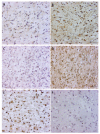
Characteristic immunohistochemical staining of active pGSK3β-Y216 and inactive pGSK3β-S9 protein in astrocytoma. (A) astrocytic brain tumor grade II with unmethylated GSK3β promoter showing weak cytoplasmic staining of pGSK3β-S9; (B) same astrocytic brain tumor grade II with unmethylated GSK3β promoter showing strong cytoplasmic and nuclear staining of pGSK3β-Y216; (C) glioblastoma (grade IV) with unmethylated GSK3β promoter showing lack of cytoplasmic staining of pGSK3β-S9; (D) same glioblastoma with unmethylated GSK3β promoter showing strong cytoplasmic and nuclear staining of pGSK3β-Y216; (E) glioblastoma (grade IV) with methylated GSK3β promoter showing moderate cytoplasmic and strong nuclear staining of pGSK3β-S9; (F) glioblastoma (grade IV) with methylated GSK3β promoter showing weak cytoplasmic staining of pGSK3β-Y216. Scale bar 50 µm.
Figure 6
Figure 6
APC exon 11/RsaI/RFLP in glioblastoma samples is demonstrated. Lane M-standard DNA 50 bp ladder (Invitrogen); lanes 1, 2: heterozygous sample (tumor and blood), both alleles, cut and uncut, are visible; lane 3: possible restriction site introduced in tumor sample; lane 4: paired homozygous sample (blood); lanes 5, 6: homozygous sample (tumor and blood), uncut alleles are visible; lanes 7, 8: homozygous sample (tumor and blood), cut alleles are visible; lane 9: LOH, cut allele is missing; lane 10: corresponding informative blood sample, both alleles, cut and uncut, are visible.

References

    1. Perry A., Wesseling P. Histologic Classification of Gliomas. Handb. Clin. Neurol. 2016;134:71–95. doi: 10.1016/B978-0-12-802997-8.00005-0. - DOI - PubMed
    1. Wesseling P., Capper D. WHO 2016 Classification of Gliomas. Neuropathol. Appl. Neurobiol. 2018;44:139–150. doi: 10.1111/nan.12432. - DOI - PubMed
    1. Louis D.N., Perry A., Reifenberger G., von Deimling A., Figarella-Branger D., Cavenee W.K., Ohgaki H., Wiestler O.D., Kleihues P., Ellison D.W. The 2016 World Health Organization Classification of Tumors of the Central Nervous System: A Summary. Acta Neuropathol. 2016;131:803–820. doi: 10.1007/s00401-016-1545-1. - DOI - PubMed
    1. Kristensen B.W., Priesterbach-Ackley L.P., Petersen J.K., Wesseling P. Molecular Pathology of Tumors of the Central Nervous System. Ann. Oncol. 2019;30:1265–1278. doi: 10.1093/annonc/mdz164. - DOI - PMC - PubMed
    1. Glibo M., Serman A., Karin-Kujundzic V., Bekavac Vlatkovic I., Miskovic B., Vranic S., Serman L. The Role of Glycogen Synthase Kinase 3 (GSK3) in Cancer with Emphasis on Ovarian Cancer Development and Progression: A Comprehensive Review. Bosn. J. Basic Med. Sci. 2021;21:5–18. doi: 10.17305/bjbms.2020.5036. - DOI - PMC - PubMed

LinkOut - more resources