Review
. 2021 May 21;22(11):5445.
doi: 10.3390/ijms22115445.

Regulation and Role of Transcription Factors in Osteogenesis

Affiliations
Review

Regulation and Role of Transcription Factors in Osteogenesis

Wilson Cheuk Wing Chan et al. Int J Mol Sci. .

Abstract

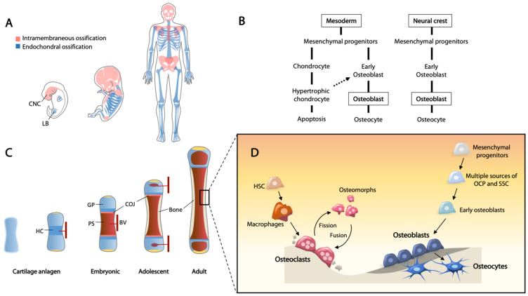
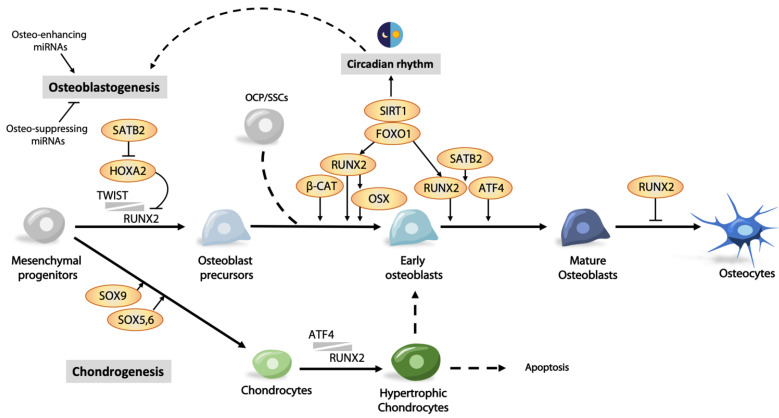
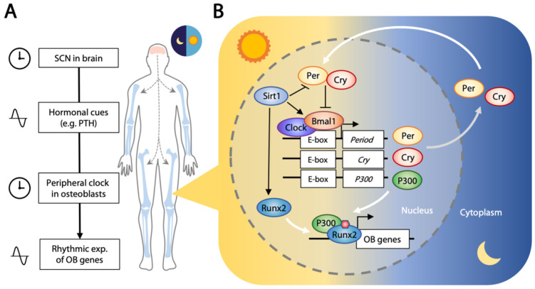
Bone is a dynamic tissue constantly responding to environmental changes such as nutritional and mechanical stress. Bone homeostasis in adult life is maintained through bone remodeling, a controlled and balanced process between bone-resorbing osteoclasts and bone-forming osteoblasts. Osteoblasts secrete matrix, with some being buried within the newly formed bone, and differentiate to osteocytes. During embryogenesis, bones are formed through intramembraneous or endochondral ossification. The former involves a direct differentiation of mesenchymal progenitor to osteoblasts, and the latter is through a cartilage template that is subsequently converted to bone. Advances in lineage tracing, cell sorting, and single-cell transcriptome studies have enabled new discoveries of gene regulation, and new populations of skeletal stem cells in multiple niches, including the cartilage growth plate, chondro-osseous junction, bone, and bone marrow, in embryonic development and postnatal life. Osteoblast differentiation is regulated by a master transcription factor RUNX2 and other factors such as OSX/SP7 and ATF4. Developmental and environmental cues affect the transcriptional activities of osteoblasts from lineage commitment to differentiation at multiple levels, fine-tuned with the involvement of co-factors, microRNAs, epigenetics, systemic factors, circadian rhythm, and the microenvironments. In this review, we will discuss these topics in relation to transcriptional controls in osteogenesis.

Keywords: bone; circadian rhythm; epigenetics; microRNA; osteoblast; osteoblast differentiation; osteogenesis; skeletogenesis; transcription factor.


Conflict of interest statement

The authors declare no conflict of interest.

Figures

Figure 1
Figure 1
Current understanding of osteogenesis and bone remodeling. (A) Flat bones are formed through intramembranous ossification, cells are originated from the cranial neural crest (CNC). Limb bones are formed through endochondral ossification (details shown in (C)), cells are originated from the mesoderm-derived limb bud (LB) mesenchyme. (B) Two major routes for osteoblast differentiation. Mesoderm cells give rise to mesenchymal osteochondroprogenitors (OCPs) which can diverge into chondrocytic and osteoblastic lineages. Chondrocytes undergo hypertrophy and a portion of them differentiate into osteoblasts at the chondro-osseous junction. Neural crest-derived mesenchymal progenitors can differentiate directly to osteoblasts during intramembraneous ossification. (C) Endochondral ossification is a process of converting cartilage to bone and is essential for bone elongation. Cartilage anlagen of a future bone forming in the limb bud during embryogenesis. Chondrocyte hypertrophy (HC) initiates in the center of the anlagen where blood vessels (BVs) invade, bringing in osteoprogenitors and bone marrow cells. The primary spongiosa (PS) separates the cartilage into proximal and distal growth plates (GPs). From childhood to adolescence, there is an active proliferation of chondrocytes prior to hypertrophy, and the mineralizing cartilage is replaced by bone at the chondro-osseous junction (COJ). Thickening of cortical bone continues from birth to puberty when the GPs become inactive. (D) Bone remodeling maintains the integrity and homeostasis of bone in adulthood. Osteoclasts are bone resorptive cells originated from hematopoietic stem cells (HSCs). They remove microfractured segments of bone and mobilize osteoblasts to form new bone. Osteomorphs are a novel cell type generated through fission of osteoclasts. Subsequent fusion of osteomorphs can reform active osteoclasts. Multiple sources of skeletal stem cells (SSCs) and OCPs have been identified as the source of osteoblasts for bone formation. Some of the mature osteoblasts are embedded into the osteoid and further differentiate into osteocytes which have a critical role in bone remodeling coordination.
Figure 2
Figure 2
Transcriptional regulation of osteoblast differentiation. SOX9 and RUNX2 are major fate determinants of mesenchymal progenitors to chondrogenesis and osteoblastogenesis, respectively. Cells can “detour” to chondrogenesis or commit to an osteoblast lineage. RUNX2 is the master transcription factor that regulates multiple steps in osteoblast commitment and differentiation. Its transcriptional activity is controlled at multiple levels such as transcriptional co-factors, inhibitors, osteo-enhancing and -suppressing miRNAs, and environmental cues such as light–dark cycle.
Figure 3
Figure 3
Circadian clock in bone. (A) Suprachiasmatic nucleus (SCN) in the hypothalamus receives the 24 h light–dark signals and conveys them in the form of nerve or hormonal signals. The rhythmic level of hormone controls the peripheral clock in bone, hence leading to rhythmic expression of osteoblastic (OB) genes. (B) The molecular clock involves the positive regulators CLOCK and BMAL1 which bind to the E-box elements and activate expression of circadian negative regulators PER and CRY. PER and CRY inhibit activities of CLOCK and BMAL1 to form a feedback loop that occurs within a period of 24 h. CLOCK/BMAL1 can bind to the E-box region and activate expression of P300 which subsequently promotes the acetylation of histone 3 and facilitates the formation of a transcriptional complex with RUNX2 to drive expression of osteoblastic genes. Sirt1 has dual roles in the circadian clock and osteogenesis. It binds CLOCK/BMAL1 in a circadian manner and promotes the deacetylation and degradation of PER and is a positive regulator of RUNX2.

References

    1. Doro D., Liu A., Grigoriadis A.E., Liu K.J. The Osteogenic Potential of the Neural Crest Lineage May Contribute to Craniosynostosis. Mol. Syndromol. 2019;10:48–57. doi: 10.1159/000493106. - DOI - PMC - PubMed
    1. Quarto N., Wan D.C., Kwan M.D., Panetta N.J., Li S., Longaker M.T. Origin matters: Differences in embryonic tissue origin and Wnt signaling determine the osteogenic potential and healing capacity of frontal and parietal calvarial bones. J. Bone Miner. Res. 2010;25:1680–1694. doi: 10.1359/jbmr.091116. - DOI - PMC - PubMed
    1. Chai Y., Maxson R.E. Recent advances in craniofacial morphogenesis. Dev. Dyn. 2006;235:2353–2375. doi: 10.1002/dvdy.20833. - DOI - PubMed
    1. Berendsen A.D., Olsen B.R. Bone development. Bone. 2015;80:14–18. doi: 10.1016/j.bone.2015.04.035. - DOI - PMC - PubMed
    1. He J., Yan J., Wang J., Zhao L., Xin Q., Zeng Y., Sun Y., Zhang H., Bai Z., Li Z., et al. Dissecting human embryonic skeletal stem cell ontogeny by single-cell transcriptomic and functional analyses. Cell Res. 2021 doi: 10.1038/s41422-021-00467-z. - DOI - PMC - PubMed

Substances

LinkOut - more resources