. 2021 May 21;22(11):5448.
doi: 10.3390/ijms22115448.

MMP-9 Deletion Attenuates Arteriovenous Fistula Neointima through Reduced Perioperative Vascular Inflammation

Affiliations

MMP-9 Deletion Attenuates Arteriovenous Fistula Neointima through Reduced Perioperative Vascular Inflammation

Yu-Chung Shih et al. Int J Mol Sci. .

Abstract

Matrix metalloproteinase 9 (MMP-9) expression is upregulated in vascular inflammation and participates in vascular remodeling, including aneurysm dilatation and arterial neointima development. Neointima at the arteriovenous (AV) fistula anastomosis site primarily causes AV fistula stenosis and failure; however, the effects of MMP-9 on perioperative AV fistula remodeling remain unknown. Therefore, we created AV fistulas (end-to-side anastomosis) in wild-type (WT) and MMP-9 knockout mice with chronic kidney disease to further clarify this. Neointima progressively developed in the AV fistula venous segment of WT mice during the four-week postoperative course, and MMP-9 knockout increased the lumen area and attenuated neointima size by reducing smooth muscle cell and collagen components. Early perioperative AV fistula mRNA sequencing data revealed that inflammation-related gene sets were negatively enriched in AV fistula of MMP-9 knockout mice compared to that in WT mice. qPCR results also showed that inflammatory genes, including tumor necrosis factor-α (TNF-α), monocyte chemoattractant protein-1 (MCP-1), interleukin-6 (IL-6), intercellular adhesion molecule-1 (ICAM-1), and vascular cell adhesion molecule-1 (VCAM-1), were downregulated. In addition, Western blot results showed that MMP-9 knockout reduced CD44 and RAC-alpha serine/threonine-protein kinase (Akt) and extracellular signal-regulated kinases (ERK) phosphorylation. In vitro, MMP-9 addition enhanced IL-6 and MCP-1 expression in vascular smooth muscle cells, as well as cell migration, which was reversed by an MMP-9 inhibitor. In conclusion, MMP-9 knockout attenuated AV fistula stenosis by reducing perioperative vascular inflammation.

Keywords: arteriovenous fistula; matrix metalloproteinase 9; neointima; vascular inflammation.


Conflict of interest statement

The authors declare no conflict of interest.

Figures

Figure 1
Figure 1
Neointima formation in the venous outflow tract of arteriovenous fistula (AVF). (A) Wild-type (WT) mice underwent 5/6 nephrectomy for chronic kidney disease (CKD) induction, followed by AVF creation four weeks later. Grossly, neointima formation was observed at proximal AVF venous segment. (B) Histologically, the initial 600 μm venous outflow tract was divided into five 100 μm thick segments as illustrated in (A), scale bar = 100 μm. After elastin staining, neointima at each segment was examined, which was found to enlarge gradually after AVF creation. **** p < 0.0001. Data presented as mean ± SEM and analyzed using one-way analysis of variance (ANOVA) followed by Bonferroni post hoc analysis; n = 6–7 in each group. AVF, arteriovenous fistula; CKD, chronic kidney disease; 2/3 Nx, 2/3 nephrectomy; UNx, uninephrectomy; WT, wild-type.
Figure 2
Figure 2
Vascular inflammation and increased matrix metalloproteinase 9 (MMP-9) expression in AVF venous segment. (A) RNA-seq data analysis revealed four out of the top 10 enriched gene ontology terms were related to inflammation. In wild-type (WT) AVF compared to contralateral veins sampled 1-week post AV fistula creation. *** p adjust < 0.001 (B) The expression of multiple proinflammatory genes, including chemokines and cytokines, was also elevated (n = 3 in each group). (C,D) Using immunohistochemical staining, MMP-9(+) cells in neointima lesions increased significantly after AV fistula surgery, scale bar = 50 μm. (n = 6–7 in each time point). (E) MMP-9 mRNA expression was upregulated at the early perioperative phase as well. (n = 6–7 in each time point). * p < 0.05, ** p < 0.01, **** p < 0.0001. Data presented as mean ± SEM and analyzed by one-way analysis of variance (ANOVA) followed by Bonferroni post hoc analysis. CTL, control; DEG, differentially expressed genes; GO, gene ontology.
Figure 3
Figure 3
Matrix metalloproteinase 9 (MMP-9) knockout attenuated neointima formation and increased lumen size in arteriovenous (AV) fistula venous segment. (A) AV fistula was created in both wild-type (WT) and MMP-9−/− mice to examine the influence on AV fistula. (B,C) Chronic kidney disease was successfully induced in both WT and MMP-9−/− mice with significantly elevated serum blood urea nitrogen (BUN) and creatinine. (D) Morphometric analysis was done after elastin staining, scale bar = 100 μm. (E) Neointima size decreased and (F) lumen area enlarged after MMP-9 knockout (p = 0.0013 and 0.0116, respectively). (G) Internal elastic lamina (IEL) perimeter was not significantly different between WT and MMP-9−/− mice. (H) IEL structure was examined under high magnification, and loose structure was found in both WT and MMP-9−/− mice AV fistula venous segment, scale bar = 50 μm. No visible detrimental effect was detected after MMP-9 knockout. * p < 0.05, ** p < 0.01. Data presented as mean ± SEM. Data analyzed by Student’s t-test one-way, n = 6–7 in each group.
Figure 3
Figure 3
Matrix metalloproteinase 9 (MMP-9) knockout attenuated neointima formation and increased lumen size in arteriovenous (AV) fistula venous segment. (A) AV fistula was created in both wild-type (WT) and MMP-9−/− mice to examine the influence on AV fistula. (B,C) Chronic kidney disease was successfully induced in both WT and MMP-9−/− mice with significantly elevated serum blood urea nitrogen (BUN) and creatinine. (D) Morphometric analysis was done after elastin staining, scale bar = 100 μm. (E) Neointima size decreased and (F) lumen area enlarged after MMP-9 knockout (p = 0.0013 and 0.0116, respectively). (G) Internal elastic lamina (IEL) perimeter was not significantly different between WT and MMP-9−/− mice. (H) IEL structure was examined under high magnification, and loose structure was found in both WT and MMP-9−/− mice AV fistula venous segment, scale bar = 50 μm. No visible detrimental effect was detected after MMP-9 knockout. * p < 0.05, ** p < 0.01. Data presented as mean ± SEM. Data analyzed by Student’s t-test one-way, n = 6–7 in each group.
Figure 4
Figure 4
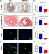
Matrix metalloproteinase 9 (MMP-9) knockout reduced α-smooth muscle actin (α-SMA) antibody and Picro Sirius stained area, and CD45(+) and Mac2(+) cells in neointima of AV fistula venous segment. (A) α-SMA(+) cells were the major cellular component within neointima. (B) The α-SMA(+) area ratio in neointima reduced after MMP-9 knockout (p < 0.0001). (C) Extracellular component of neointima was assessed with Picro Sirius Red staining. (D) The Picro Sirius Red(+) area ratio within neointima decreased in MMP-9−/− mice (p < 0.0001). (E) Leukocyte infiltration within neointima was determined by CD45 immunofluorescence. (F) MMP-9 deletion decreased CD45(+) cells/HPF in neointima (p < 0.0001). (G) Macrophages in the neointima was examined by Mac2 immunofluorescence. (H) The number of Mac2(+) cells/HPF decreased in MMP-9−/− mice (p = 0.0011). White arrows indicated positively stained cells. ** p < 0.01, **** p < 0.0001. Data presented as mean ± SEM. Data analyzed by Student’s t-test one-way, n = 6–7 in each group. HPF, high power field.
Figure 5
Figure 5
Genome-wide mRNA expression difference of WT and MMP-9−/− mice AV fistulas. (A) Gene set enrichment analysis (GSEA) with Hallmark gene sets was performed to analyze the differentially expressed genes between WT and MMP-9−/− mice. Significantly enriched pathways in different categories are visualized using bubble plots of normalized enrichment score (NES) for different comparisons, including WT AVF vs. WT CTL and MMP9 AVF vs. WT AVF, and the size of each bubble is proportional to the number of core genes within the pathway. Only pathways with FDR < 0.25 were included. TNFA_SIGNALING_VIA_NFKB was positively enriched in WT AVF vs. WT CTL, but negatively enriched in MMP9 AVF vs. WT AVF. (B) GSEA enrichment plots of immune-related pathways in MMP-9−/− AVF vs. WT AVF. (C) Leading-edge analysis of six immune-related pathways. (D) The normalized expression value of these 14 key driver genes were expressed in heatmap, n = 3 in each group. Expression of inflammation-related genes usually associated with AV fistula stenosis such as (E) TNF-α, (F) IL-6, (G) MCP-1, (H) ICAM-1, and (I) VCAM-1, were examined by qPCR, n = 6–7 in each group. * p < 0.05, ** p < 0.01, **** p < 0.0001. Data presented as mean ± SEM. Data analyzed by one-way analysis of variance (ANOVA) followed by Bonferroni post hoc analysis. ICAM-1, intercellular adhesion molecule 1; IL-6, interleukin 6; MCP-1, monocyte chemoattractant protein 1; NES, normalized enrichment score; NFKB, nuclear factor kappa-light-chain-enhancer of activated B cells; TNFA and TNF-α, tumor necrosis factor-α; VCAM-1, vascular cell adhesion molecule 1.
Figure 6
Figure 6
MMP-2 mRNA expression in AV fistula venous segment. (A) MMP-2 mRNA expression elevated after AV fistula creation comparing to contralateral veins sampled at 1 week, 2 weeks, and 4 weeks after AV fistula creation and the peak was at 2 weeks (n = 6~7 in each time point). (B,C) The deletion of MMP-9 did not make compensatory elevation of MMP-2 at 1-week and 4-weeks post AVF creation. (n = 6–7 in each group). * p < 0.05, **** p < 0.0001. Data presented as mean ± SEM and analyzed by one-way ANOVA followed by Bonferroni post hoc analysis for multiple comparison and Student’s t-test for comparison of two.
Figure 7
Figure 7
Matrix metalloproteinase 9 (MMP-9) knockout downregulated CD44 and RAC-alpha serine/threonine-protein kinase (Akt) and extracellular signal-regulated kinases (ERK) phosphorylation in arteriovenous (AV) fistula venous segment. (A) Western blotting was done for evaluating the protein expression in AV fistula venous segment. (B) One-week post AV fistula creation, CD44 expression was increased in wild-type (WT) mice, which was reversed by MMP-9 knockout. (C) Akt phosphorylation was also increased after AV fistula creation and attenuated by MMP-9 knockout. (D) Phospho-ERK level was significantly decreased in MMP-9−/− mice. *** p < 0.001, **** p < 0.0001. Data presented as mean ± SEM. Data analyzed by one-way analysis of variance (ANOVA) followed by Bonferroni post hoc analysis, n = 6 in each group. CTL, control.
Figure 8
Figure 8
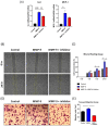
Mouse vascular smooth muscle cell line (MOVAS) was used for in vitro study. (A) MCP-1 and IL-6 mRNA expression was assessed by qPCR. The influence of matrix metalloproteinase 9 (MMP-9) on MOVAS migration was assessed using (B,C) wound healing and (D,E) transwell migration assays. * p < 0.05, ** p< 0.01, *** p < 0.001, **** p < 0.0001. Data presented as mean ± SEM. Data analyzed by one-way analysis of variance (ANOVA) followed by Bonferroni post hoc analysis, n = 6 in each group.

Similar articles

Cited by

References

    1. Sands J.J. Increasing AV fistulas: Revisiting a time-tested solution. Semin. Dial. 2000;13:351–353. doi: 10.1046/j.1525-139x.2000.00098.x. - DOI - PubMed
    1. Roy-Chaudhury P., Lee T.C. Vascular stenosis: Biology and interventions. Curr. Opin. Nephrol. Hypertens. 2007;16:516–522. doi: 10.1097/MNH.0b013e3282efa57f. - DOI - PubMed
    1. Rooijens P.P., Tordoir J.H., Stijnen T., Burgmans J.P., Smet de A.A., Yo T.I. Radiocephalic wrist arteriovenous fistula for hemodialysis: Meta-analysis indicates a high primary failure rate. Eur. J. Vasc. Endovasc. Surg. 2004;28:583–589. doi: 10.1016/j.ejvs.2004.08.014. - DOI - PubMed
    1. Huijbregts H.J., Bots M.L., Wittens C.H., Schrama Y.C., Moll F.L., Blankestijn P.J., CIMINO Study Group Hemodialysis arteriovenous fistula patency revisited: Results of a prospective, multicenter initiative. Clin. J. Am. Soc. Nephrol. 2008;3:714–719. doi: 10.2215/CJN.02950707. - DOI - PMC - PubMed
    1. Al-Jaishi A.A., Oliver M.J., Thomas S.M., Lok C.E., Zhang J.C., Garg A.X., Kosa S.D., Quinn R.R., Moist L.M. Patency rates of the arteriovenous fistula for hemodialysis: A systematic review and meta-analysis. Am. J. Kidney Dis. 2014;63:464–478. doi: 10.1053/j.ajkd.2013.08.023. - DOI - PubMed

Substances

LinkOut - more resources