Review
. 2021 May 21;11(6):775.
doi: 10.3390/biom11060775.

Anti-Angiogenic Property of Free Human Oligosaccharides

Affiliations
Review

Anti-Angiogenic Property of Free Human Oligosaccharides

Boram Bae et al. Biomolecules. .

Abstract

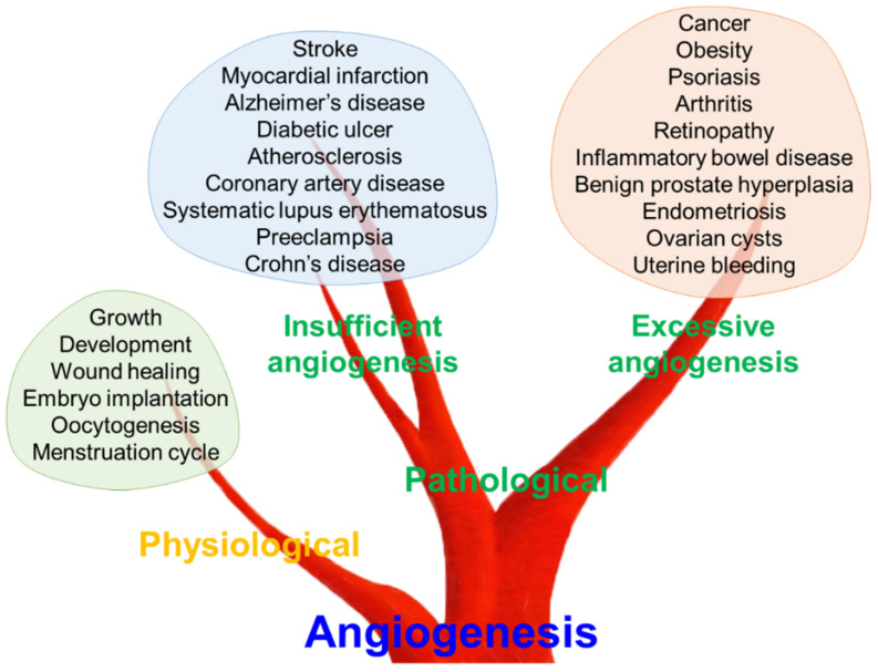
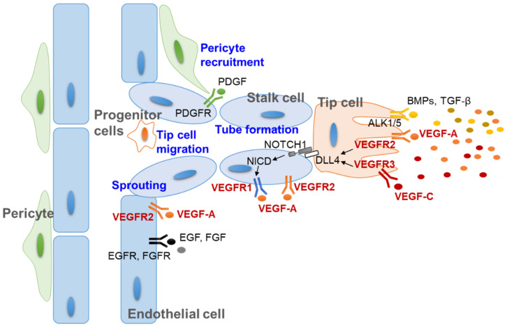
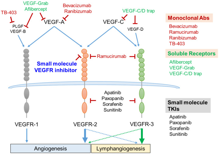
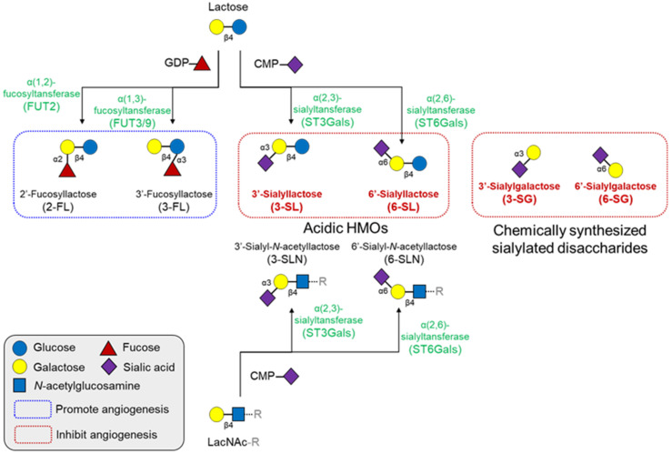
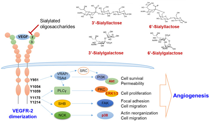
Angiogenesis, a fundamental process in human physiology and pathology, has attracted considerable attention owing to its potential as a therapeutic strategy. Vascular endothelial growth factor (VEGF) and its receptor (VEGFR) are deemed major mediators of angiogenesis. To date, inhibition of the VEGF-A/VEGFR-2 axis has been an effective strategy employed in the development of anticancer drugs. However, some limitations, such as low efficacy and side effects, need to be addressed. Several drug candidates have been discovered, including small molecule compounds, recombinant proteins, and oligosaccharides. In this review, we focus on human oligosaccharides as modulators of angiogenesis. In particular, sialylated human milk oligosaccharides (HMOs) play a significant role in the inhibition of VEGFR-2-mediated angiogenesis. We discuss the structural features concerning the interaction between sialylated HMOs and VEGFR-2 as a molecular mechanism of anti-angiogenesis modulation and its effectiveness in vivo experiments. In the current state, extensive clinical trials are required to develop a novel VEGFR-2 inhibitor from sialylated HMOs.

Keywords: VEGFR-2; angiogenesis; human milk oligosaccharides; inhibitor; sialyllactose.


Conflict of interest statement

The authors declare no conflict of interest.

Figures

Figure 1
Figure 1
Role of angiogenesis in physiological and pathological conditions. Angiogenesis is a fundamental process in human physiology and pathology. Dysregulated angiogenesis, both insufficient and excessive angiogenesis, can lead to various pathological conditions.
Figure 2
Figure 2
Molecular mechanism of angiogenesis and multicellular interaction during new vessel development. In response to stimulators such as VEGFs, vascular endothelial cells sprout from the basement membrane and migrate to the site of new vessel formation. The formation of tip and stalk cells is regulated by the VEGF-DLL4/NOTCH signaling pathway. The other stimulating molecules, such as PDGF, EGF, FGF, BMPs, and TGF-β, also cooperate to regulate pericytes and tip cells. ALK1/5, actin receptor-like kinase 1/5; BMP, bone morphogenic protein (yellow dots); DLL4, delta-like protein 4; EGF, epidermal growth factor (black dot); EGFR, epidermal growth factor receptor; FGF, fibroblast growth factor (gray dot); FGFR, fibroblast growth factor receptor; NICD, intracellular domain of the notch protein; PDGF, platelet-derived growth factor (green dot); PDGFR, platelet-derived growth factor receptor; TGF-β, transforming growth factor-β (dark yellow dots); VEGF-A, vascular endothelial cell growth factor A (orange dots); VEGF-C, vascular endothelial cell growth factor C (red dots); VEGFR, vascular endothelial cell growth factor receptor.
Figure 3
Figure 3
VEGF/VEGFR axis as a therapeutic target and its intervention strategies. Five members of the VEGF family are composed of VEGF-A, VEGF-B, VEGF-C, VEGF-D, and PLGF. VEGF-A binds to both VEGFR-1 and VEGFR-2. VEGF-C binds to both VEGFR-2 and VEGFR-3. VEGF-B and PLGF bind to VEGFR-1, and VEGF-D binds to VEGFR-3. VEGFR-1- and VEGFR-2-mediated signaling cascades regulate vascular angiogenesis. VEGFR-3 activation is essential for lymphangiogenesis (green arrow). VEGFR-2 cooperatively activates lymphangiogenesis with VEGFR-3 (blue dashed arrow), and VEGFR-3 also slightly enhances the vessel angiogenesis (green dashed arrow). Several monoclonal antibodies and recombinant soluble receptors consisting of the extracellular domains of VEGFRs have been successfully developed as therapeutic anti-angiogenic agents. Small molecules targeting the intracellular tyrosine kinase domain or extracellular VEGF-binding domain are under development as novel strategies for inhibiting angiogenesis. Abs, antibodies; PLGF, placenta growth factor; TKIs, tyrosine kinase inhibitors; VEGF, vascular endothelial cell growth factor; VEGFR, vascular endothelial cell growth factor receptor.
Figure 4
Figure 4
Structures and synthetic pathways of major HMOs and their effects on angiogenesis. All HMOs consist of a lactose core or LacNAc core, with a few exceptions. These cores can be enzymatically elongated in repeats of LacNAc. The elongated HMO chains can be further decorated with fucosylation or sialylation by fucosyltransferases or sialyltransferases, respectively. Fucosylated HMOs generally promotes angiogenesis, but several sialyllactose analogs inhibit angiogenesis. CMP, cytidine monophosphate; GDP, guanosine diphosphate; HMO, human milk oligosaccharides; LacNAc, N-acetyllactosamine.
Figure 5
Figure 5
Molecular mechanism of the anti-angiogenic action by sialic acid-containing oligosaccharides. Sialylated lactose and sialylgalactose with sialic acid linked α2-3 or α2-6 to galactose inhibit the activation of VEGFR-2 by interfering the binding between VEGF and VEGFR-2 via Ig-like domain 2 and 3 (green circle). Inhibition of VEGFR-2 activation thereby suppresses the downstream angiogenic signaling pathways, such as the PI3K/Akt, PKC/ERK1/2 and p38 pathways. ERK1/2, extracellular signaling-regulated kinases 1/2; FAK, focal adhesion kinase; NCK, non-catalytic region of tyrosine kinase; PI3K, phosphoinositide 3-kinase; PLCγ, phospholipase γ; PKC, protein kinase C; SHB, SH2 domain-containing adapter protein B; TSAd, T-cell specific adaptor protein; VEGF, vascular endothelial cell growth factor; VEGFR, vascular endothelial cell growth factor receptor; VRAP, VEGF-receptor activated protein.

References

    1. Hill D.R., Newburg D.S. Clinical applications of bioactive milk components. Nutr. Rev. 2015;73:463–476. doi: 10.1093/nutrit/nuv009. - DOI - PMC - PubMed
    1. Ballard O., Morrow A.L. Human milk composition: Nutrients and bioactive factors. Pediatr. Clin. N. Am. 2013;60:49–74. doi: 10.1016/j.pcl.2012.10.002. - DOI - PMC - PubMed
    1. Andreas N.J., Kampmann B., Mehring Le-Doare K. Human breast milk: A review on its composition and bioactivity. Early Hum. Dev. 2015;91:629–635. doi: 10.1016/j.earlhumdev.2015.08.013. - DOI - PubMed
    1. Bode L. Human milk oligosaccharides: Every baby needs a sugar mama. Glycobiology. 2012;22:1147–1162. doi: 10.1093/glycob/cws074. - DOI - PMC - PubMed
    1. Thurl S., Munzert M., Boehm G., Matthews C., Stahl B. Systematic review of the concentrations of oligosaccharides in human milk. Nutr. Rev. 2017;75:920–933. doi: 10.1093/nutrit/nux044. - DOI - PMC - PubMed

Publication types

MeSH terms

Substances

LinkOut - more resources