Review
. 2021 May 21;10(6):1283.
doi: 10.3390/cells10061283.

Major Depression: One Brain, One Disease, One Set of Intertwined Processes

Affiliations
Review

Major Depression: One Brain, One Disease, One Set of Intertwined Processes

Elena V Filatova et al. Cells. .

Abstract

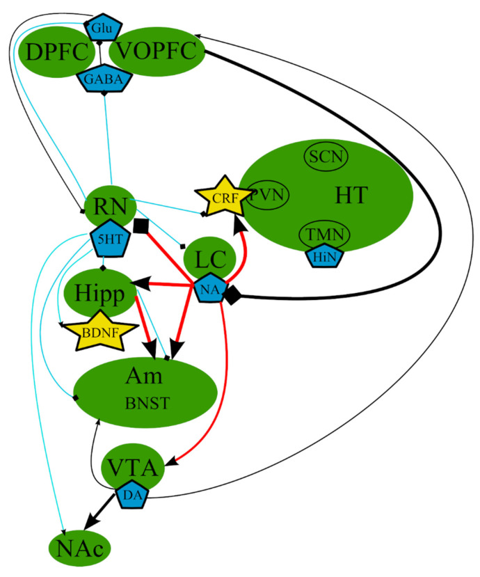
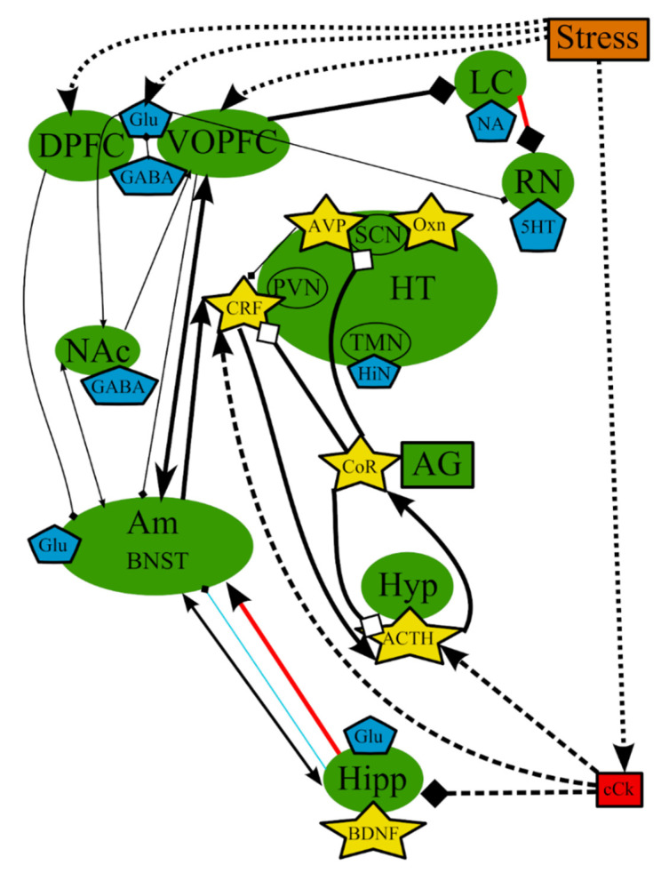
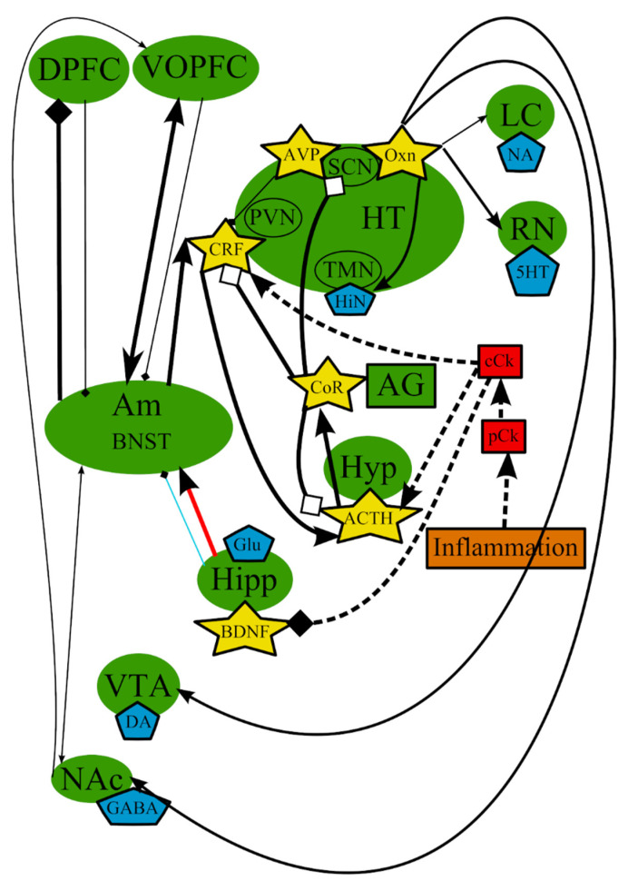
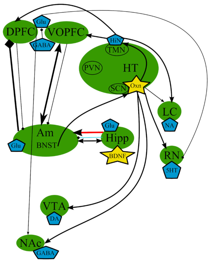
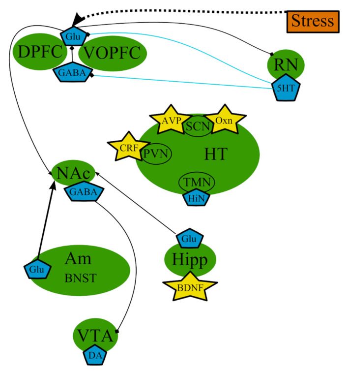
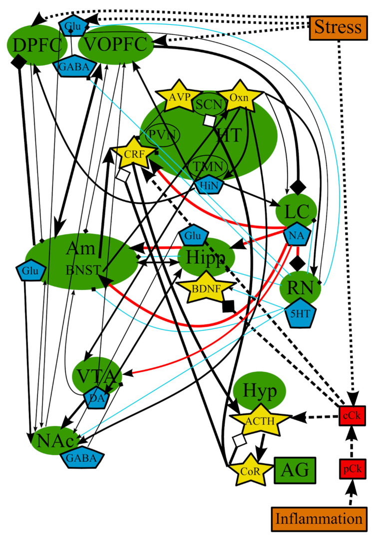
Major depressive disorder (MDD) is a heterogeneous disease affecting one out of five individuals and is the leading cause of disability worldwide. Presently, MDD is considered a multifactorial disease with various causes such as genetic susceptibility, stress, and other pathological processes. Multiple studies allowed the formulation of several theories attempting to describe the development of MDD. However, none of these hypotheses are comprehensive because none of them can explain all cases, mechanisms, and symptoms of MDD. Nevertheless, all of these theories share some common pathways, which lead us to believe that these hypotheses depict several pieces of the same big puzzle. Therefore, in this review, we provide a brief description of these theories and their strengths and weaknesses in an attempt to highlight the common mechanisms and relationships of all major theories of depression and combine them together to present the current overall picture. The analysis of all hypotheses suggests that there is interdependence between all the brain structures and various substances involved in the pathogenesis of MDD, which could be not entirely universal, but can affect all of the brain regions, to one degree or another, depending on the triggering factor, which, in turn, could explain the different subtypes of MDD.

Keywords: common mechanisms; etiology; major depressive disorder; pathogenesis; theories of depression.


Conflict of interest statement

The authors declare no conflict of interest.

Figures

Figure 1
Figure 1
The monoamine hypothesis: main brain structures, neurotransmitters, biologically active substances, and interactions. BDNF—brain-derived neurotrophic factor; BNST—bed nuclei stria terminalis; CRF—corticotropin-releasing factor; DA—dopamine; DPFC—dorsal prefrontal cortex; Glu—glutamine; GABA—gamma-aminobutyric acid; HiN—histamine; Hipp—hippocampus; HT—hypothalamus; 5-HT—serotonin; LC—locus ceruleus; NA—noradrenaline; NAc—nucleus accumbens; Oxn—orexin; SCN—suprachiasmatic nucleus; TMN—tuberomammilar nucleus; RN—raphe nucleus; PVN—paraventricular nucleus; VOPFC—ventral and orbital prefrontal cortex; VTA—ventral tegmental area; -> (arrow): activating effect; -<> (rhombus): a black rhombus—inhibitory effect; thick line—effect is increased; thin line—effect is decreased; medium thickness line—effect is not changed, or alterations of the effect are unknown; red line—noradrenaline effect; blue line—serotonin effect; black line—various neurotransmitters or neuropeptides.
Figure 2
Figure 2
The hypothesis of stress-induced depression: main structures, neurotransmitters, biologically active substances, interactions, and external factors. ACTH—adrenocorticotropic hormone; AG—adrenal gland; Am—amygdala; AVP—arginine-vasopressin; BDNF—brain-derived neurotrophic factor; BNST—bed nuclei stria terminalis; cCk—central pro-inflammatory cytokines; CoR—cortisol; CRF—corticotropin-releasing factor; DA—dopamine; DPFC—dorsal prefrontal cortex; Glu—glutamine; GABA—gamma-aminobutyric acid; HiN—histamine; Hipp—hippocampus; HT—hypothalamus; 5-HT—serotonin; Hyp—hypophysis; LC—locus ceruleus; NA—noradrenaline; NAc—nucleus accumbens; Oxn—orexin; SCN—suprachiasmatic nucleus; TMN—tuberomammilar nucleus; RN—raphe nucleus; PVN—paraventricular nucleus; VOPFC—ventral and orbital prefrontal cortex; -> (arrow): activating effect; -<> (rhombus): a black rhombus—inhibitory effect; a white rhombus: an effect is blocked or ineffective because the receptor is not sensitive; thick line—effect is increased; thin line—effect is decreased; medium thickness line—effect is not changed or alterations of the effect are unknown; red line—noradrenaline effect (most of them were omitted to simplify the figure); blue line—serotonin effect (most of them were omitted to simplify the figure); black line—various neurotransmitters or neuropeptides; dotted lines—influence of external factors.
Figure 3
Figure 3
The inflammation/cytokine hypothesis: main structures, neurotransmitters, biologically active substances, interactions, and external factors. ACTH—adrenocorticotropic hormone; AG—adrenal gland; Am—amygdala; AVP—arginine-vasopressin; BDNF—brain-derived neurotrophic factor; BNST—bed nuclei stria terminalis; cCk—central pro-inflammatory cytokines; CoR—cortisol; CRF—corticotropin-releasing factor; DA—dopamine; DPFC—dorsal prefrontal cortex; Glu—glutamine; GABA—gamma-aminobutyric acid; HiN—histamine; Hipp—hippocampus; HT—hypothalamus; 5-HT—serotonin; Hyp—hypophysis; LC—locus ceruleus; NA—noradrenaline; NAc—nucleus accumbens; Oxn—orexin; SCN—suprachiasmatic nucleus; TMN—tuberomammilar nucleus; RN—raphe nucleus; pCk—peripheral pro-inflammatory cytokines; PVN—paraventricular nucleus; VOPFC—ventral and orbital prefrontal cortex; VTA—ventral tegmental area; -> (arrow): activating effect; -<> (rhombus): a black rhombus—inhibitory effect; a white rhombus: an effect is blocked or ineffective because the receptor is not sensitive; thick line—effect is increased; thin line—effect is decreased; medium thickness line—effect is not changed or alterations of the effect are unknown; red line—noradrenaline effect (most of them were omitted to simplify the figure); blue line—serotonin effect (most of them were omitted to simplify the figure); black line—various neurotransmitters or neuropeptides; dotted lines—influence of external factors.
Figure 4
Figure 4
The circadian hypothesis: main brain structures, neurotransmitters, biologically active substances, and interactions. Am—amygdala; AVP—arginine-vasopressin; BDNF—brain-derived neurotrophic factor; BNST—bed nuclei stria terminalis; CRF—corticotropin-releasing factor; DA—dopamine; DPFC—dorsal prefrontal cortex; Glu—glutamine; GABA—gamma-aminobutyric acid; HiN—histamine; Hipp—hippocampus; HT—serotonin; Hyp—hypophysis; LC—locus ceruleus; NA—noradrenaline; NAc—nucleus accumbens; Oxn—orexin; SCN—suprachiasmatic nucleus; TMN—tuberomammilar nucleus; RN—raphe nucleus; PVN—paraventricular nucleus; VOPFC—ventral and orbital prefrontal cortex; VTA—ventral tegmental area; -> (arrow): activating effect; -<> (rhombus): a black rhombus—inhibitory effect; thick line—effect is increased; thin line—effect is decreased; medium thickness line—effect is not changed or alterations of the effect are unknown; red line—noradrenaline effect (most of them were omitted to simplify the figure); blue line—serotonin effect (most of them were omitted to simplify the figure); black line—various neurotransmitters or neuropeptides.
Figure 5
Figure 5
The excitatory neurotransmitters involved in the pathogenesis of MDD: main brain structures, neurotransmitters, biologically active substances, interactions, and external factors. Am—amygdala; AVP—arginine-vasopressin; BDNF—brain-derived neurotrophic factor; BNST—bed nuclei stria terminalis; CRF—corticotropin-releasing factor; DA—dopamine; DPFC—dorsal prefrontal cortex; Glu—glutamine; GABA—gamma-aminobutyric acid; HiN—histamine; Hipp—hippocampus; HT—hypothalamus; 5-HT—serotonin; NA—noradrenaline; NAc—nucleus accumbens; Oxn—orexin; SCN—suprachiasmatic nucleus; TMN—tuberomammilar nucleus; RN—raphe nucleus; PVN—paraventricular nucleus; VOPFC—ventral and orbital prefrontal cortex; VTA—ventral tegmental area; -> (arrow): activating effect; -<> (rhombus): a black rhombus—inhibitory effect; thick line—the effect is increased; thin line—the effect is decreased; medium thickness line—the effect is not changed or alterations of the effect are unknown; blue line—serotonin effect (most of them were omitted to simplify the figure); black line—various neurotransmitters or neuropeptides.
Figure 6
Figure 6
The overall picture of the combined common mechanisms and relationships of all major theories of depression. ACTH—adrenocorticotropic hormone; AG—adrenal gland; Am—amygdala; AVP—arginine-vasopressin; BDNF—brain-derived neurotrophic factor; BNST—bed nuclei stria terminalis; cCk—central pro-inflammatory cytokines; CoR—cortisol; CRF—corticotropin-releasing factor; DA—dopamine; DPFC—dorsal prefrontal cortex; Glu—glutamine; GABA—gamma-aminobutyric acid; HiN—histamine; Hipp—hippocampus; HT—hypothalamus; 5-HT—serotonin; Hyp—hypophysis; LC—locus ceruleus; NA—noradrenaline; NAc—nucleus accumbens; Oxn—orexin; SCN—suprachiasmatic nucleus; TMN—tuberomammilar nucleus; RN—raphe nucleus; pCk—peripheral pro-inflammatory cytokines; PVN—paraventricular nucleus; VOPFC—ventral and orbital prefrontal cortex; VTA—ventral tegmental area; -> (arrow): activating effect; -<> (rhombus): a black rhombus—inhibitory effect; a white rhombus—an effect is blocked or ineffective because the receptor is not sensitive; thick line—effect is increased; thin line—effect is decreased; medium thickness line—the effect is not changed or alterations of the effect are unknown; red line—noradrenaline effect; blue line—serotonin effect; black line—various neurotransmitters or neuropeptides; dotted lines—influence of external factors.

Similar articles

Cited by

References

    1. Menard C., Hodes G.E., Russo S.J. Pathogenesis of depression: Insights from human and rodent studies. Neuroscience. 2016;321:138–162. doi: 10.1016/j.neuroscience.2015.05.053. - DOI - PMC - PubMed
    1. Russo S.J., Nestler E.J. The brain reward circuitry in mood disorders. Nat. Rev. Neurosci. 2013;14:609–625. doi: 10.1038/nrn3381. - DOI - PMC - PubMed
    1. Krishnan V., Nestler E.J. The molecular neurobiology of depression. Nature. 2008;455:894–902. doi: 10.1038/nature07455. - DOI - PMC - PubMed
    1. Schmidt H.D., Shelton R.C., Duman R.S. Functional biomarkers of depression: Diagnosis, treatment, and pathophysiology. Neuropsychopharmacology. 2011;36:2375–2394. doi: 10.1038/npp.2011.151. - DOI - PMC - PubMed
    1. Shadrina M., Bondarenko E.A., Slominsky P.A. Genetics Factors in Major Depression Disease. Front. Psychiatry. 2018;9:334. doi: 10.3389/fpsyt.2018.00334. - DOI - PMC - PubMed

Publication types

MeSH terms

LinkOut - more resources