. 2021 May 4;11(5):416.
doi: 10.3390/life11050416.

Molecular Insights into the Genetic Variability of ORF Virus in a Mediterranean Region (Sardinia, Italy)

Affiliations

Molecular Insights into the Genetic Variability of ORF Virus in a Mediterranean Region (Sardinia, Italy)

Elisabetta Coradduzza et al. Life (Basel). .

Abstract

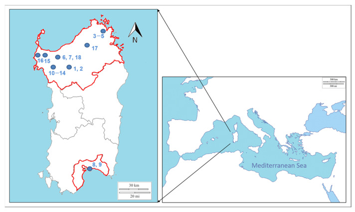
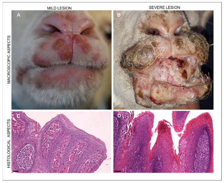
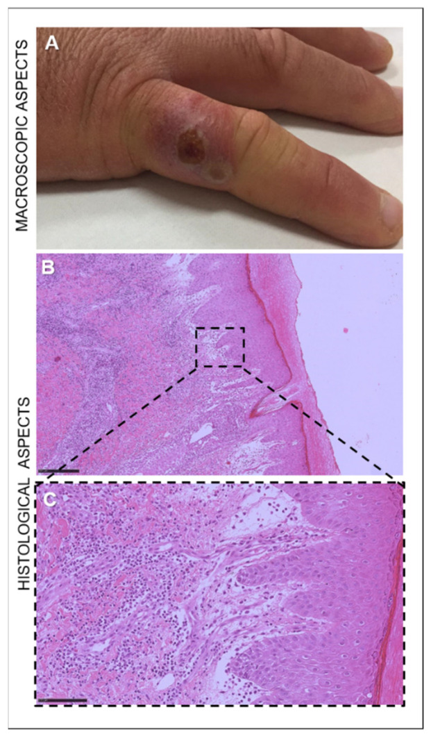
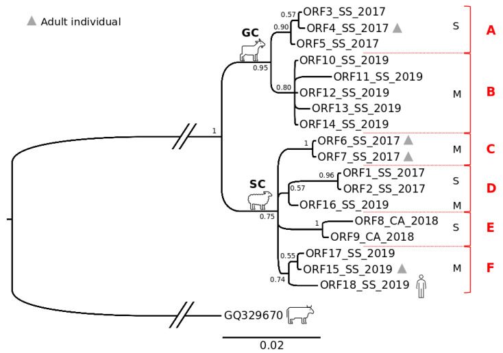
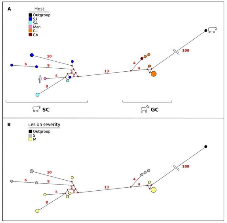
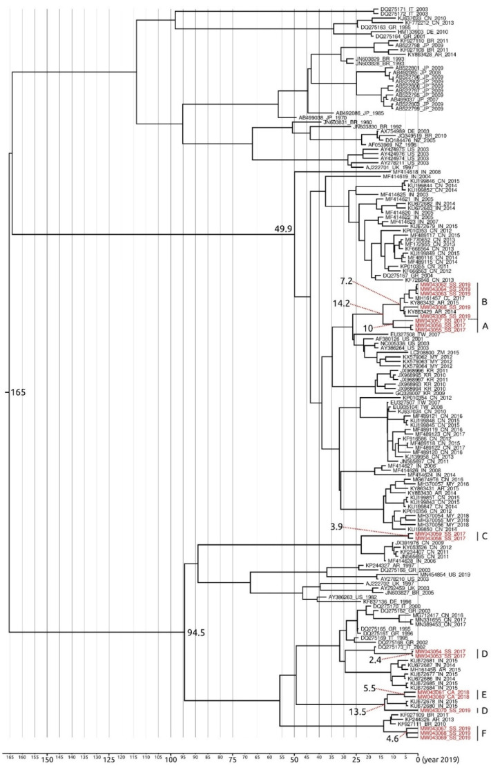
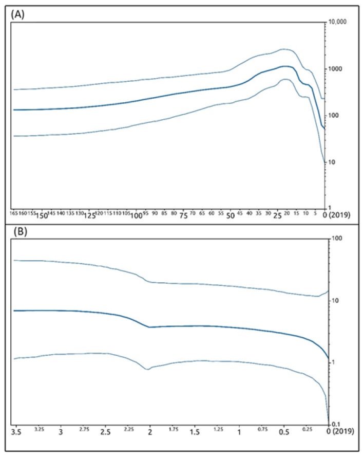
Orf virus (ORFV) represents the causative agent of contagious ecthyma, clinically characterized by mild papular and pustular to severe proliferative lesions, mainly occurring in sheep and goats. In order to provide hints on the evolutionary history of this virus, we carried out a study aimed to assess the genetic variation of ORFV in Sardinia that hosts a large affected small ruminant population. We also found a high worldwide mutational viral evolutionary rate, which resulted, in turn, higher than the rate we detected for the strains isolated in Sardinia. In addition, a well-supported genetic divergence was found between the viral strains isolated from sheep and those from goats, but no relevant connection was evidenced between the severity of lesions produced by ORFV and specific polymorphic patterns in the two species of hosts. Such a finding suggests that ORFV infection-related lesions are not necessarily linked to the expression of one of the three genes here analyzed and could rather be the effect of the expression of other genes or rather represents a multifactorial character.

Keywords: B2L; O45; VIR; contagious ecthyma; molecular dating; phylogenetic analysis; sequencing.


Conflict of interest statement

The authors and the founders declare no conflict of interest.

Figures

Figure 1
Figure 1
Sampling plan. The geographic locations of the farms where samples were collected are indicated in the map of the Mediterranean island of Sardinia. The numbers at each site refer to the sample codes as reported in Table 1. The hyphen between two numbers indicates an interval. Blue spots indicate the municipalities where the farms were located, as reported in Table 1.
Figure 2
Figure 2
Representative images of the macroscopic appearance of ORFV-infected lambs and microscopical patterns of ORFV-infected lambs with mild and severe lesions. (A) Mild lesions in an ORFV-infected lamb showing vesicles and crusted pustules in the upper and lower lips. (B) Severe lesions appearing in an ORFV-affected lamb as coalescing hyperemic, proliferative, verrucous outgrowths with a papilloma-like appearance covering the incisor tooth, present in the lips together with crusted lesions. (C) Mild lesions histologically showing moderate epithelial hyperplasia, hyperkeratosis with elongated rete ridges, and proliferations of neovascular structures in the dermis of the host skin. Hematoxylin and eosin staining (H&E). Original magnification 100×. Scale bar = 100 µm. (D) Severe manifestation of ORFV-infected animals microscopically characterized by massive proliferative patterns involving the epithelium and showing hyperkeratosis and hyperplastic epithelium with extremely elongated rete ridges and ballooning degeneration. In the lamina propria of the buccal mucosae and in the derma of skin, there is evident proliferation of mesenchymal cells resembling hemangiomatous patterns. Hematoxylin and eosin staining (H&E). Original magnification 100×. Scale bar = 100 µm.
Figure 3
Figure 3
Nodule on the right little finger of a 30-year-old sheep farmer infected with ORFV. (A) Targetoid nodule with a necrotic center, a white ring, and peripheral erythema. (B) Microscopical appearance of the skin of the ORF-infected man, characterized by marked acanthosis and hyperkeratosis. Hematoxylin and eosin staining (H&E). Original magnification 50×. Scale bar = 250 µm. (C) In the dermis, vascular proliferation with granulocytic and eosinophilic infiltration. Hematoxylin and eosin staining (H&E). Original magnification 400×. Scale bar = 100 µm.
Figure 4
Figure 4
ORFV CPE resulting from infection by scab homogenate on VERO cells. (A) mock; (B) early stage; (C) intermediate state; (D) final stage.
Figure 5
Figure 5
Bayesian phylogenetic tree based on the Sardinian ORFV concatenated gene dataset (B2L-VIR-O45). Values of node supports are expressed in posterior probabilities. The sample codes are as reported in Table 1. S: severe pattern of lesions; M: mild pattern of lesions. Red capital letters on the right indicate the genetic sub-groups described in the text.
Figure 6
Figure 6
Median-joining network based on the Sardinian ORFV concatenated dataset (B2L-VIR-O45). (A) The haplotype spots in the graphic are colored according to host species (sheep vs. goats). SJ: lamb; SA: ewe; GJ: kid; GA: goat. (B) The haplotype spots in the graphic are colored according to the type of lesions showed by hosts. S: severe pattern of lesion; M: mild pattern of lesion. The small red plots on the nodes show median vectors representing the hypothetical connecting sequences that were calculated using the maximum parsimony method. Only the numbers of mutations between haplotypes that are greater than 1 are reported on network branches.
Figure 7
Figure 7
Bayesian phylogenetic time-scaled maximum clade credibility tree based on the ORFV VIR global dataset. All main nodes of the tree are highly supported by values of posterior probabilities ≥0.95. Nodes with a percentage of posterior probability <0.95 were considered to be not statistically supported. The Sardinian samples codes are reported in red font (further details in the supplementary Table S1). The codes of sequences from GenBank report the accession number, the country acronym, and the presumptive year of isolation. The numbers at the nodes refer to the dating (in years before 2019) of the cluster. Capital letters on the right refer to the genetic sub-groups indicated in Figure 5. Horizontal bar indicates the years before 2019 in a temporal scale.
Figure 8
Figure 8
Bayesian skyline plots. Bayesian skyline plots depicting the evolutionary history of ORFV VIR gene. The effective population size of viral infection (y-axis) is shown as a function of time (x-axis), with the units expressed as years. (A) VIR global dataset; (B) VIR regional dataset encompassing only Sardinian sequences. The bold lines denote the median effective number of infections and the thin lines (placed above and below the median value line) demarcate the 95% highest posterior density (HPD) confidence interval.

References

    1. Bergqvist C., Kurban M., Abbas O. Orf virus infection. Rev. Med. Virol. 2017;27:1–9. doi: 10.1002/rmv.1932. - DOI - PubMed
    1. Hosamani M., Scagliarini A., Bhanuprakash V., McInnes C.J., Singh R.K. Orf: An update on current research and future perspectives. Expert Rev. Anti Infect. Ther. 2009;7:879–893. doi: 10.1586/eri.09.64. - DOI - PubMed
    1. Vaccari F. Ph.D. Thesis. University of Bologna; Bologna, Italy: 2009. Evolutionary Mechanisms of Parapoxviruses: Genomic Characterization of Pseudocowpoxviruses and Development of Systems for the Study of Recombinations.
    1. Zeller H. South-West African Goat Pox. Arb. Reichsgesundheitsamte. 1920;52:501–537.
    1. Kumar R., Trivedi R.N., Bhatt P., Khan S.H., Khurana S.K., Tiwari R., Karthik K., Malik Y.S., Dhama K., Chandra R. Contagious pustular dermatitis (orf disease)—Epidemiology, diagnosis, control and public health concerns. Adv. Anim. Vet. Sci. 2015;3:649–676. doi: 10.14737/journal.aavs/2015/3.12.649.676. - DOI

LinkOut - more resources