. 2021 May 4;11(5):374.
doi: 10.3390/jpm11050374.

In Situ Maturated Early-Stage Human-Induced Pluripotent Stem Cell-Derived Cardiomyocytes Improve Cardiac Function by Enhancing Segmental Contraction in Infarcted Rats

Affiliations

In Situ Maturated Early-Stage Human-Induced Pluripotent Stem Cell-Derived Cardiomyocytes Improve Cardiac Function by Enhancing Segmental Contraction in Infarcted Rats

Diogo Biagi et al. J Pers Med. .

Abstract

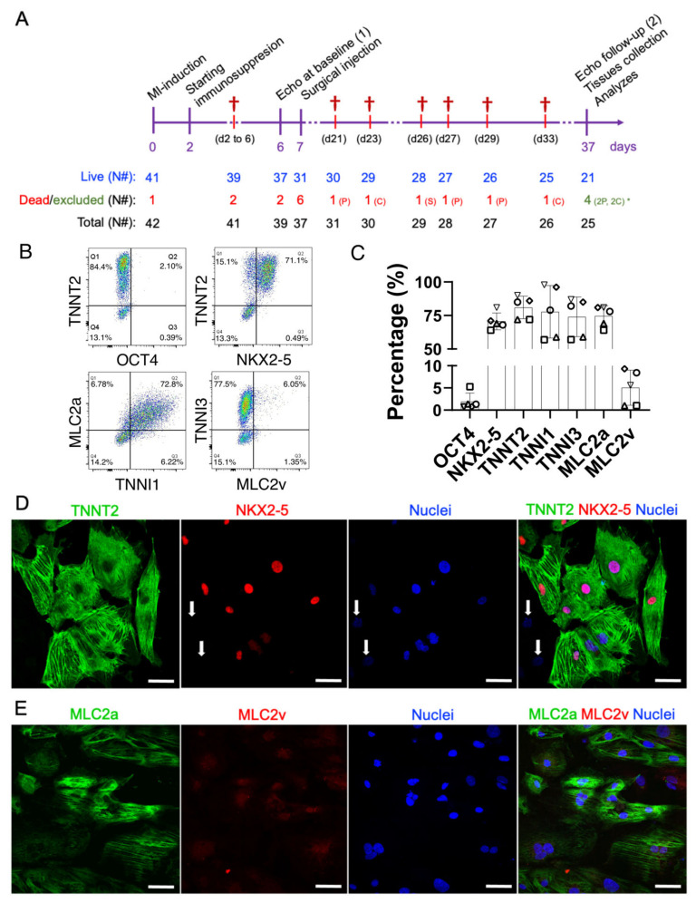
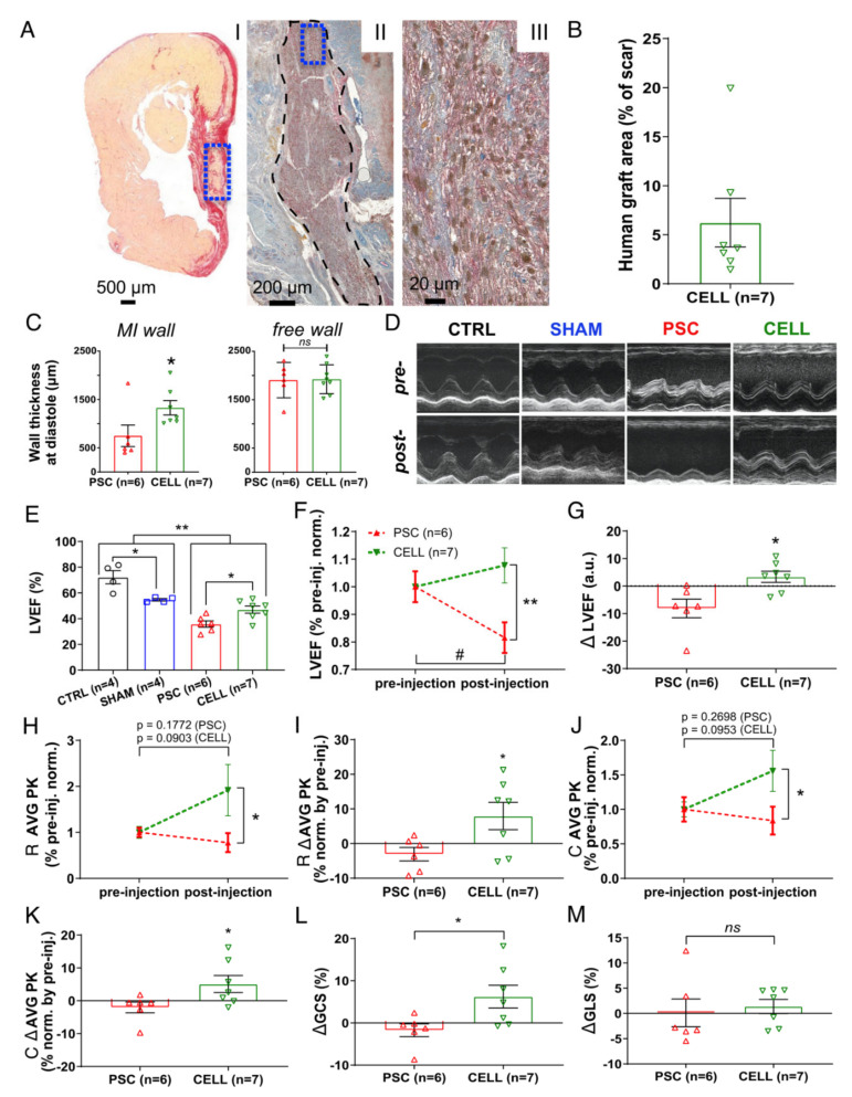
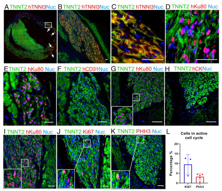
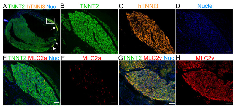
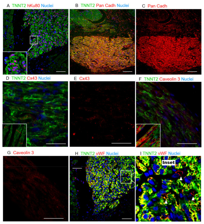
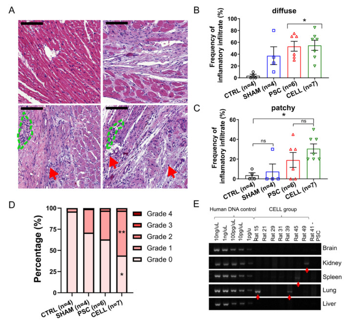
The scant ability of cardiomyocytes to proliferate makes heart regeneration one of the biggest challenges of science. Current therapies do not contemplate heart re-muscularization. In this scenario, stem cell-based approaches have been proposed to overcome this lack of regeneration. We hypothesize that early-stage hiPSC-derived cardiomyocytes (hiPSC-CMs) could enhance the cardiac function of rats after myocardial infarction (MI). Animals were subjected to the permanent occlusion of the left ventricle (LV) anterior descending coronary artery (LAD). Seven days after MI, early-stage hiPSC-CMs were injected intramyocardially. Rats were subjected to echocardiography pre-and post-treatment. Thirty days after the injections were administered, treated rats displayed 6.2% human cardiac grafts, which were characterized molecularly. Left ventricle ejection fraction (LVEF) was improved by 7.8% in cell-injected rats, while placebo controls showed an 18.2% deterioration. Additionally, cell-treated rats displayed a 92% and 56% increase in radial and circumferential strains, respectively. Human cardiac grafts maturate in situ, preserving proliferation with 10% Ki67 and 3% PHH3 positive nuclei. Grafts were perfused by host vasculature with no evidence for immune rejection nor ectopic tissue formations. Our findings support the use of early-stage hiPSC-CMs as an alternative therapy to treat MI. The next steps of preclinical development include efficacy studies in large animals on the path to clinical-grade regenerative therapy targeting human patients.

Keywords: cardiac function; cardiomyocytes; heart failure; human induced pluripotent stem cells; myocardial infarction; regeneration; stem cell-therapy.


Conflict of interest statement

The authors declared the following potential conflicts of interest concerning the research, authorship, and/or publication of this article: The authors D.B., E.T.F., J.C.C.O., M.V.N., A.F.R.Jr., S.R., I.O., R.V., M.V., E.C., and R.D were employees of PluriCell Biotech during the period in which the study was conducted. D.B., M.V., E.C., and R.D. own shares in PluriCell Biotech (D.B. and M.V. are co-founders). The other authors report no conflicts.

Figures

Figure 1
Figure 1
Therapeutic study design and molecular characterization of the early-stage hiPSC-CMs. (A) On day zero, myocardial infarctions were induced by the permanent occlusion of the proximal AD coronary artery. Forty-eight hours after MI (day 2), the immunosuppression protocol was started. CsA was administered twice a day intraperitoneally until the end of the protocol (day 37). Six days after the MI induction (day 6), all animals were subjected to baseline echocardiography and randomized. On day 7, animals from placebo (pro-survival cocktail—PSC) and CELL groups were subjected to a second surgical thoracotomy followed by the intramyocardial injection of hiPSC-CMs. Thirty days after injection (37 days after the MI induction), all survivors were subjected to a final echocardiography and were then euthanized, necropsied, and had their hearts and other organs collected and stored for further analysis. (B) Representative scatterplots of the injected hiPSC-CM population on days 11–15 of differentiation. (C) The number of positive cells expressing cardiac markers such as TNNT2, TNNI1, TNNI3, MLC2A, MLC2V, and NKX2-5, and a pluripotency marker OCT4, was obtained by flow cytometry, expressed as a percentage, and plotted in bar charts (mean ± SD). (D) Representative immunofluorescence demonstrating the expression of cardiac markers in hiPSC-CMs on day 13 of differentiation (TNNT2, NKX2-5, and nuclei). Except for two cells in the image (white arrows), all the TNNT2-positive cells were also NKX2-5 positive. (E) MLC2a, MLC2v, nuclei, and merged image. Note that the expression of MLC2v seems to be nuclear/perinuclear, strongly corroborating the lack of maturation of early-stage hiPSC-CMs. Scale bars: 100 µm.
Figure 2
Figure 2
Early-stage hiPSC-CM-based therapy improves segmental contraction resulting in overall LV cardiac function preservation on infarcted rats. Echocardiography was performed on days 6 (1 day prior to injections) and 37 (30 days after injection) to evaluate the cell therapy effects. (A) Representative panel of histological images from animal 29 (CELL group). (AI) Papillary level cross-section stained with picrosirius red evidencing “live” cardiac tissue (yellow) and fibrosis (red). “Live” cardiac tissue was found in the scar (blue box). (AII) Subsequent cross-section subjected to an immunohistochemical reaction against human Ku80 (brown nuclei) and Troponin T2 (red), evidencing human cells in the rat heart (black dashed area). (AIII) The human cardiac graft is taken from positive cells for both markers (brown nuclei—hKu80, and red fibers—Troponin T2) (From blue box in AII). (B) Percentage of the area covered by hKu80-positive cells in the scar tissue was quantified in 3–5 cross-sections at the papillary level. The human graft areas were expressed as a percentage relative to the scar area. All the CELL-treated rats displayed hKu80/Troponin T2-positive areas. (C) Wall thickness at diastole was measured at the papillary level using low magnification images. MI (left chart) and free wall (right chart) values were calculated as a mean of three measurements covering the whole scar extension and expressed as μm. (D) Representative short-axis of M-mode images pre- (day 6) and post-injection (day 37). (E) LVEF was calculated using short-axis images at four LV depths by a modified Simpson algorithm. CELL-treated rats showed improved LVEF 30 days post-treatment (vs. PSC, * p < 0.05). Furthermore, MI-induced animals showed significantly lower LVEF values than the CTRL and SHAM groups (** p < 0.01). Additionally, the CTRL and SHAM groups displayed different LVEF values (* p < 0.05). (F) The LVEF values of PSC and CELL rats were normalized by their respective pre-treatment values and plotted over time. CELL-treated rats show higher LVEF values than PSCs post-treatment (** p < 0.01). PSCs showed a significant deterioration of LVEF 37 days after MI-induction (# p = 0.0145), whereas CELL rats had their LVEF preserved (p = 0.2162). (G) The difference in LVEF values between pre- and post-injection (delta) shows cardiac function preservation in the CELL group (* p = 0.0117 vs. PSC). (H) Radial strain time-to-peak (AVG PK) of PSC and CELL rats were normalized by their respective pre-treatment values. The CELL group displayed higher radial strain values AVG PK post-treatment (* p = 0.0441). Pre- vs. post-treatment analysis revealed no significant differences in the PSC nor CELL group, as demonstrated by the p-values printed in the image. (I) Radial strain time-to-peak values between pre- and post-injection (delta) were also significantly different (* p < 0.0371). (J) Circumferential strain time-to-peak (AVG PK) values of the PSC and CELL rats were normalized by their respective pre-treatment values. The CELL group displayed higher circumferential strain AVG PK post-treatment (* p = 0.0494). Pre- vs. post-treatment analysis revealed no significant differences in the PSC nor CELL group, as demonstrated by the p-values printed in the image. (K) The circumferential strain time-to-peak delta was also significantly different (* p = 0.0465). (L) The global circumferential strain between pre- and post-injection (delta) was significantly positive for CELL-treated animals (p = 0.0324 vs. PSC), but (M) the global longitudinal strain between pre- and post-injection (delta) did not display changes (data positively normalized; p = 0.6767 CELL vs. PSC). Data showed as mean ± SE. R: radial; C: circumferential; GCS: global circumferential strain; GLS: global longitudinal strain.
Figure 3
Figure 3
Representative image from a human graft showing cardiac cellular composition. (A) Panoramic immunofluorescence staining for TNNT2 (green, rat and human) and human TNNI3 (red, human-specific): arrows indicate grafts. (B) An amplified view of largest graft region marked by the white rectangle in A. (C) Higher magnification of (B) showing sarcomere structure organization. (D) Image showing that hiPSC-CMs follow cardiac structure organization, TNNT2 (green) and hKu80 (pink) by overlap for hKu80 (red) and nuclei (blue). (EH) Images showing no evidence of hiPSC-CMs trans-differentiation into cell types other than cardiomyocytes. Serial slices were labeled for hKu80 (E) and human CD31 (F). The same strategy was used to label hKu80 (G) and human Cytokeratin-1 (H). (IL) Ki67 and PHH3 markers evidenced that the human cardiac grafts were composed of hiPSC-CMs with an active cell cycle. (I) Serial slides were labeled as TNNT2 (green) and hKu80 (pink), (J) TNNT2 (green) and Ki67 (pink), and (K) TNNT2 (green) and PHH3 (pink). Pink represents the overlap of red and blue (nuclei). (L) Quantification for cell-cycle markers presents a percentage of Ki67-positive cells and PHH3-positive cells (vs. the total number of hKu80-positive nuclei for each image analyzed). Ki67: n = 3 and PHH3: n = 5. Scale bars: 50 µm.
Figure 4
Figure 4
Immunofluorescence staining for MLC2a and MLC2v 30 days after cell transplantation demonstrating the maturation of hTNNI3-positive cells in vivo. (A) Panoramic immunofluorescence staining for TNNT2 (human and rat cardiomyocytes) and hTNNI3 (human-specific cardiomyocytes). (BD) Images showing the area depicted by the white rectangle in A (hTNNI3-positive cardiomyocytes) in higher magnification with a single channel for each marker. (B) TNNT2 (green, rat and human cells), (C) human TNNI3 (orange, note the absence of rat tissue compared with (B,D) nuclei (blue). (E) Serial slices were stained with TNNT2 (green), MLC2a (red), and nuclei (blue). Note that the graft in C is the same as that used in E. (F) MLC2a (red) showing scant expression on the graft. (G) Merged image for TNNT2 (green), MLC2v (red), and nuclei (blue). (H) MLC2v showing extensive labeling, compared with MLC2a in (F). Scale bars: 100 µm.
Figure 5
Figure 5
Histological evaluation of hiPSC-CM grafts suggesting limited host/graft interaction. (A) Colocalization of hKu80 (red) and nuclei (blue), and TNNT2 (green). (B) Pan-cadherin expression in both the host and human grafts indicates a high cell-to-cell interaction. (C) Pan-cadherin is more highly expressed on the human cardiac graft than on the host tissue. (D) The expression of Cx43 is evidently lower and more randomly distributed across the human cardiac cells, whereas a structured expression can be observed in the host tissue (inset), particularly on the short-borders of adult cardiomyocytes. (E) Cx43 single-channel image. (F) Caveolin3 (red) was also found in grafts at lower levels of expression and organization than in adult cardiomyocytes, where this protein is structurally expressed inside the caveolae within the T-tubules (inset), suggesting hiPSC-CMs limited maturation. (G) Cav3 single-channel image. (H,I) Rat vWF-positive blood vessels (red) perfusing the human cardiac grafts, observed in abundance in all the animals tested. (I) A magnification of H: white arrowheads show individual vWF-positive vessels (red stain). Scale bars: 50 µm.
Figure 6
Figure 6
Immune-rejection against human cardiac grafts and the biodistribution of hiPSC-CMs over the body. Pathological assessments were performed through H&E-stained slides to quantify the incidence, distribution, and severity of inflammatory infiltrates. Representative images of heart sections for (A) CTRL, SHAM, PSC, and CELL rats (left to right, top to bottom). Red arrows indicate sparse diffuse inflammatory cells, and the blue dashed lines indicate small patches of inflammatory cells (scale bar: 100 µm). The frequency of (B) diffuse and (C) patchy infiltrates was quantitatively measured and expressed as a percentage. (D) The severity of the inflammatory infiltrate was estimated using a well-established grade system by observing the size and spread of inflammatory cells in the sections assessed. Grades were transformed into a percentage of severity per image and therefore per animal and were plotted as stacked percentage bars. Grade 2 areas were observed only in infarcted animals. Grades 0 and 1 estimations were significantly different between the CTRL and CELL groups (* p < 0.05, and ** p < 0.01, respectively). (E) PCR results for human mitochondrial DNA indicate the presence of human material in rat 15 (lung), rat 39 (lung), rat 45 (spleen), and rat 49 (kidney), shown by the red arrows.

References

    1. Teo K.K., Rafiq T. Cardiovascular risk factors and prevention: A perspective from developing countries. Can. J. Cardiol. 2021 doi: 10.1016/j.cjca.2021.02.009. - DOI - PubMed
    1. Virani S.S., Alonso A., Aparicio H.J., Benjamin E.J., Bittencourt M.S., Callaway C.W., Carson A.P., Chamberlain A.M., Cheng S., Delling F.N., et al. Heart Disease and Stroke Statistics—2021 Update. Circulation. 2021;143 doi: 10.1161/CIR.0000000000000950. - DOI - PubMed
    1. Porrello E.R., Mahmoud A.I., Simpson E., Hill J.A., Richardson J.A., Olson E.N., Sadek H.A. Transient regenerative potential of the neonatal mouse heart. Science. 2011;331:1078–1080. doi: 10.1126/science.1200708. - DOI - PMC - PubMed
    1. Vivien C.J., Hudson J.E., Porrello E.R. Evolution, comparative biology and ontogeny of vertebrate heart regeneration. NPJ Regen. Med. 2016;1:16012. doi: 10.1038/npjregenmed.2016.12. - DOI - PMC - PubMed
    1. Mummery C.L., Davis R.P., Krieger J.E. Challenges in using stem cells for cardiac repair. Sci. Transl. Med. 2010;2:27ps17. doi: 10.1126/scitranslmed.3000558. - DOI - PubMed

LinkOut - more resources