. 2021 May 4;10(9):1974.
doi: 10.3390/jcm10091974.

The Spectrum of COVID-19-Associated Myocarditis: A Patient-Tailored Multidisciplinary Approach

Affiliations

The Spectrum of COVID-19-Associated Myocarditis: A Patient-Tailored Multidisciplinary Approach

Giovanni Peretto et al. J Clin Med. .

Abstract

Background: Myocarditis lacks systematic characterization in COVID-19 patients.

Methods: We enrolled consecutive patients with newly diagnosed myocarditis in the context of COVID-19 infection. Diagnostic and treatment strategies were driven by a dedicated multidisciplinary disease unit for myocarditis. Multimodal outcomes were assessed during prospective follow-up.

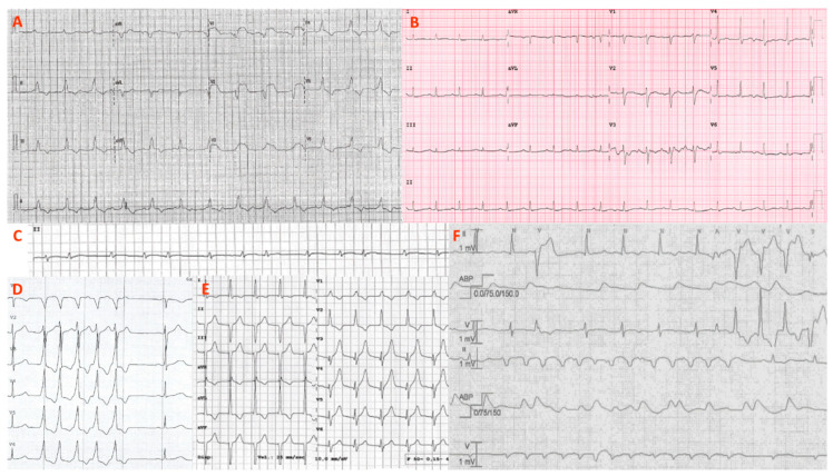
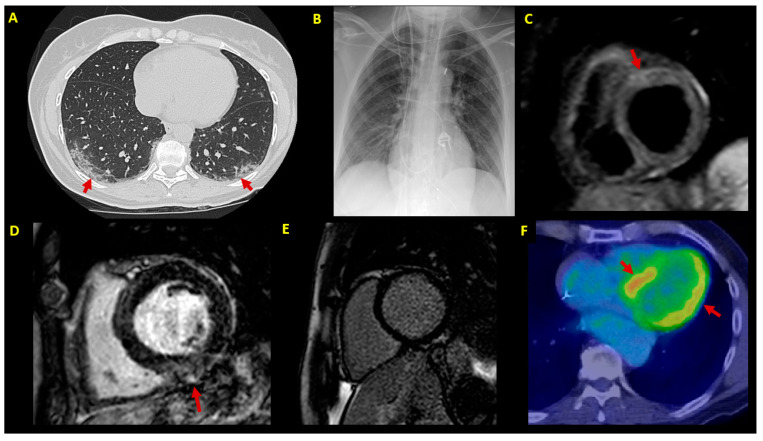
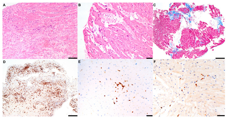
Results: Seven consecutive patients (57% males, age 51 ± 9 y) with acute COVID-19 infection received a de novo diagnosis of myocarditis. Endomyocardial biopsy was of choice in hemodynamically unstable patients (n = 4, mean left ventricular ejection fraction (LVEF) 25 ± 9%), whereas cardiac magnetic resonance constituted the first exam in stable patients (n = 3, mean LVEF 48 ± 10%). Polymerase chain reaction (PCR) analysis revealed an intra-myocardial SARS-CoV-2 genome in one of the six cases undergoing biopsy: in the remaining patients, myocarditis was either due to other viruses (n = 2) or virus-negative (n = 3). Hemodynamic support was needed for four unstable patients (57%), whereas a cardiac device implant was chosen in two of four cases showing ventricular arrhythmias. Medical treatment included immunosuppression (43%) and biological therapy (29%). By the 6-month median follow-up, no patient died or experienced malignant arrhythmias. However, two cases (29%) were screened for heart transplantation.

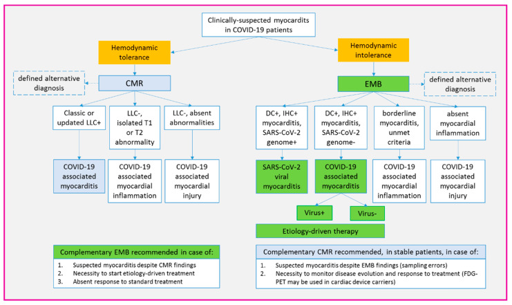
Conclusions: Myocarditis associated with acute COVID-19 infection is a spectrum of clinical manifestations and underlying etiologies. A multidisciplinary approach is the cornerstone for tailored management.

Keywords: COVID-19; SARS-CoV-2; cardiac magnetic resonance; endomyocardial biopsy; immunosuppression; inflammation; multidisciplinary; myocarditis; ventricular arrhythmias.


Conflict of interest statement

The authors declare no conflict of interest.

Figures

Figure 1
Figure 1
ECG and arrhythmias. Representative ECGs and arrhythmias from the patient series (P1–P7) are shown. (A) Atrioventricular dissociation with huge anterior ST elevation (P7); (B) sinus tachycardia with low QRS voltages and diffuse repolarization abnormalities (P2); (C) paroxysmal atrial fibrillation detected by ICU telemonitoring (P7); (D) nonsustained ventricular tachycardia (P4); (E) transitory, self-limited, accelerated junctional rhythm (P1); (F) polymorphic and irregular nonsustained ventricular tachycardias during ICU stay (P5). ICU = intensive care unit.
Figure 2
Figure 2
Imaging findings. Imaging findings at patient (P1–P7) diagnostic workup. (A) Chest CT scan showing bilateral patchy ground-glass opacities (arrows) (P3); (B) chest X-ray in a patient (P7) with cardiogenic shock supported by IABP, VA-ECMO and temporary pacemaker; (C) CMR in a patient with infarct-like acute myocarditis associated with COVID-19 (P3); T2-STIR sequence shows edema in the anterior basal segment (arrow); (D) LGE sequences in a patient (P5) showing mild inferior mid-myocardial/subepicardial LGE (arrow); (E) absence of LGE by 3-month follow-up CMR in a patient (P4) with fulminant myocarditis at presentation; (F) 3-month follow-up FDG-PET scan in an ICD carrier (P5) with virus-negative myocarditis; abnormal left ventricular FDG uptake is shown (arrows). CMR = cardiac magnetic resonance; CT = computed tomography; EMB = endomyocardial biopsy; FDG-PET= 18F-fluorodeoxyglucose positron emission tomography; IABP = intra-aortic balloon pump; ICD = implantable cardioverter defibrillator; LGE = late gadolinium enhancement; LVEF = left ventricular ejection fraction; STIR = short-tau inversion recovery; VA-ECMO = venoarterial extracorporeal membrane oxygenator.
Figure 3
Figure 3
Histology findings. Representative findings at histologic analysis of patients (P1–P7) are shown. (A) Hematoxylin-eosin assay in a patient (P7) with fulminant EBV myocarditis complicating COVID-19 infection; diffuse areas of necrosis and massive lymphocytic inflammatory infiltrates are shown; (B) mild lymphocytic infiltration without significant necrosis in a patient (P1) with borderline myocarditis; (C) trichrome assay in a patient (P6) with myocarditis (not shown) and likely preexisting dilated cardiomyopathy; multiple areas of replacement fibrosis (blue) are shown; (D) immunohistochemistry for CD3+ T-lymphocytes (>7/mm2) in fulminant myocarditis (P7); (E) in contrast, milder CD3+ infiltrated in a patient with mild inflammatory cardiomyopathy (P5); (F) CD68+ macrophage infiltrates in a patient with SARS-CoV-2 borderline myocarditis (P2). Scale bars: (A): 100 µm; (B,E,F): 50 µm; (C,D): 200 µm. CD = cluster of differentiation; EBV = Epstein–Barr virus; SARS-CoV2 = severe acute respiratory syndrome coronavirus-2.
Figure 4
Figure 4
Proposed algorithm for diagnosis and classification of myocarditis in COVID-19 patients. Proposed diagnostic workup for patients with clinically suspected myocarditis associated with COVID-19 infection. The key criterion to guide the initial choice between CMR and EMB in hemodynamic tolerance. However, when feasible, we suggest an extensive use of both techniques, since they provide complementary information. Remarkably, based on the current definition of viral myocarditis (2,6), only EMB allows for SARS-CoV-2 to be identified as responsible for myocardial inflammation. In general, etiology-driven therapy is safe only when myocarditis diagnosis is EMB-proven. CMR = cardiac magnetic resonance; DC = Dallas criteria; EMB = endomyocardial biopsy; FDG-PET= 18F-fluorodeoxyglucose positron emission tomography; IHC = immunohistochemistry; LLC = Lake Louise criteria; SARS-CoV-2 = severe acute respiratory syndrome coronavirus-2.

References

    1. Hendren N.S., Drazner M.H., Bozkurt B., Cooper L.T., Jr. Description and Proposed Management of the Acute COVID-19 Cardiovascular Syndrome. Circulation. 2020;141:1903–1914. doi: 10.1161/CIRCULATIONAHA.120.047349. - DOI - PMC - PubMed
    1. Tschöpe C., Ammirati E., Bozkurt B., Caforio A.L.P., Cooper L.T., Felix S.B., Hare J.M., Heidecker B., Heymans S., Hübner N., et al. Myocarditis and inflammatory cardiomyopathy: Current evidence and future directions. Nat. Rev. Cardiol. 2020:1–25. doi: 10.1038/s41569-020-00435-x. - DOI - PMC - PubMed
    1. Peretto G., Sala S., Caforio A.L.P. Acute myocardial injury, MINOCA, or myocarditis? Improving characterization of coronavirus-associated myocardial involvement. Eur. Heart J. 2020;41:2124–2125. doi: 10.1093/eurheartj/ehaa396. - DOI - PMC - PubMed
    1. Peretto G., De Luca G., Campochiaro C., Palmisano A., Busnardo E., Sartorelli S., Barzaghi F., Cicalese M.P., Esposito A., Sala S. Telemedicine in myocarditis: Evolution of a mutidisciplinary "disease unit" at the time of COVID-19 pandemic. Am. Heart J. 2020;229:121–126. doi: 10.1016/j.ahj.2020.07.015. - DOI - PMC - PubMed
    1. Ferreira V.M., Schulz-Menger J., Holmvang G., Kramer C.M., Carbone I., Sechtem U., Kindermann I., Gutberlet M., Cooper L.T., Liu P., et al. Cardiovascular Magnetic Resonance in Nonischemic Myocardial Inflammation: Expert Recommendations. J. Am. Coll. Cardiol. 2018;72:3158–3176. doi: 10.1016/j.jacc.2018.09.072. - DOI - PubMed

LinkOut - more resources