. 2021 May 4;10(9):1976.
doi: 10.3390/jcm10091976.

Production of IL-31 in CD45RO+CLA+H4R+ T Cells in Atopic Dermatitis

Affiliations

Production of IL-31 in CD45RO+CLA+H4R+ T Cells in Atopic Dermatitis

Chul Hwan Bang et al. J Clin Med. .

Abstract

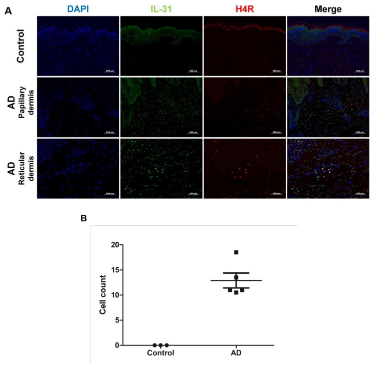
IL-31 is involved in pruritus in atopic dermatitis (AD) and the pathogenesis of AD. However, the mechanism of IL-31 production is not fully understood. We sought to investigate the association between CD45RO+CLA+H4R+ T cells and IL-31 production. Immunofluorescence studies were performed retrospectively on punch-biopsy specimens from five people with AD and three healthy controls. In addition, blood samples were collected prospectively from eight patients with AD and eight healthy controls for sorting CD45RO+CLA+H4R+ T cells. There was no overlap of patients between the biopsy group and the blood sampling group. Sorted cells were stimulated with 4-methylhistamine (4MH), and the level of IL-31 was measured by an enzyme-linked immunosorbent assay. Immunofluorescence showed co-localization of H4R and IL-31 in lesional AD skin but not in normal skin of healthy controls. The proportion of CLA+H4R+ T cells among CD3+CD45RO+ lymphocytes was 18.3 ± 6.2% in patients with AD and 11.2 ± 7.6% in healthy controls. In the AD group, production of IL-31 by CD45RO+CLA+H4R+ T cells increased from 32.4 ± 13.3 pg/mL to 47.5 ± 18.7 pg/mL by 4MH stimulation after 24 h (p < 0.001). However, in the control group, production of IL-31 was 20.1 ± 10.6 pg/mL without and 22.1 ± 9.3 pg/mL with 4MH stimulation (p > 0.05). According to our study, CD45RO+CLA+H4R+ T cells are an important source of IL-31 in AD, and may be a target for treatment of IL-31-induced pruritus.

Keywords: Interleukin-31; atopic dermatitis; histamine-4-receptor.


Conflict of interest statement

The authors declare no conflict of interest.

Figures

Figure 1
Figure 1
Co-localization of IL-31 and H4R in AD and healthy controls. (A) Overlay (yellow) of IL-31+ (green) and H4R+ (red) in AD samples and healthy controls. DAPI (blue) was used to counterstain nuclei. Original magnification × 200, scale bar = 100 μm for control and reticular dermis of AD. Original magnification × 100, scale bar = 200 μm for papillary dermis of AD. (B) IL-31+ and H4R+ co-localized cell counts in healthy controls and patients with AD. High-power field, × 200. Error bars indicate SEMs.
Figure 2
Figure 2
Result of CD45RO+CLA+H4R+ T cells in AD and healthy controls. The FACS results of CD45RO+CLA+H4R+ T cells in (A) healthy controls and (B) patients with AD. (C) Proportion of CLA+H4R+ T cells in CD3+CD45RO+ T cells; 9.1 ± 5.6% in patients with AD and 4.8 ± 3.8% in healthy control. AD, atopic dermatitis. FACS, fluorescence-activated cell sorting.
Figure 3
Figure 3
Simultaneous expression of IL-31 and H4R in sorted CD45RO+CLA+H4R+ T cells of AD patients with 4-methylhistamine (4MH) stimulation. Isolated CD45RO+CLA+H4R+ T cells from healthy controls and AD samples were cultured and stimulated with or without 4MH at 10 μM. Immunofluorescence images of IL-31 (green) and H4R (red) in sorted CD45RO+CLA+H4R+ T cells were observed with original magnification × 400, scale bar = 50 μm.
Figure 4
Figure 4
The production of IL-31 in CD45RO+CLA+H4R+ T cells and CD45RO+CLA+H4R T cells. CD45RO+CLA+H4R T cells and CD45RO+CLA+H4R+ T cells were isolated from blood samples acquired from healthy controls and patients with AD. These sorted cells were cultured at a density of 1.5 × 10⁴ cells/well and stimulated with or without 4MH at 10 μM. After stimulation for 24 h, supernatant harvested from cell medium was evaluated by ELISA for level of IL-31. * p < 0.05, *** p < 0.001.

References

    1. Dillon S.R., Sprecher C., Hammond A., Bilsborough J., Rosenfeld-Franklin M., Presnell S.R., Haugen H.S., Maurer M., Harder B., Johnston J., et al. Interleukin 31, a cytokine produced by activated T cells, induces dermatitis in mice. Nat. Immunol. 2004;5:752–760. doi: 10.1038/ni1084. - DOI - PubMed
    1. Bagci I.S., Ruzicka T. IL-31: A new key player in dermatology and beyond. J. Allergy Clin. Immunol. 2018;141:858–866. doi: 10.1016/j.jaci.2017.10.045. - DOI - PubMed
    1. Ezzat M.H., Hasan Z.E., Shaheen K.Y. Serum measurement of interleukin-31 (IL-31) in paediatric atopic dermatitis: Elevated levels correlate with severity scoring. J. Eur. Acad. Dermatol. Venereol. 2011;25:334–339. doi: 10.1111/j.1468-3083.2010.03794.x. - DOI - PubMed
    1. Ruzicka T., Hanifin J.M., Furue M., Pulka G., Mlynarczyk I., Wollenberg A., Galus R., Etoh T., Mihara R., Yoshida H., et al. Anti-Interleukin-31 Receptor A Antibody for Atopic Dermatitis. N. Engl. J. Med. 2017;376:826–835. doi: 10.1056/NEJMoa1606490. - DOI - PubMed
    1. Bilsborough J., Leung D.Y., Maurer M., Howell M., Boguniewicz M., Yao L., Storey H., LeCiel C., Harder B., Gross J.A. IL-31 is associated with cutaneous lymphocyte antigen-positive skin homing T cells in patients with atopic dermatitis. J. Allergy Clin. Immunol. 2006;117:418–425. doi: 10.1016/j.jaci.2005.10.046. - DOI - PubMed