Review
. 2021 May 11;13(10):2287.
doi: 10.3390/cancers13102287.

AKT in Bone Metastasis of Solid Tumors: A Comprehensive Review

Affiliations
Review

AKT in Bone Metastasis of Solid Tumors: A Comprehensive Review

Nico Hinz et al. Cancers (Basel). .

Abstract

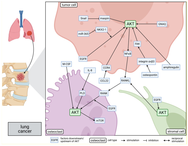
Solid tumors, such as breast cancer and prostate cancer, often form bone metastases in the course of the disease. Patients with bone metastases frequently develop complications, such as pathological fractures or hypercalcemia and exhibit a reduced life expectancy. Thus, it is of vital importance to improve the treatment of bone metastases. A possible approach is to target signaling pathways, such as the PI3K/AKT pathway, which is frequently dysregulated in solid tumors. Therefore, we sought to review the role of the serine/threonine kinase AKT in bone metastasis. In general, activation of AKT signaling was shown to be associated with the formation of bone metastases from solid tumors. More precisely, AKT gets activated in tumor cells by a plethora of bone-derived growth factors and cytokines. Subsequently, AKT promotes the bone-metastatic capacities of tumor cells through distinct signaling pathways and secretion of bone cell-stimulating factors. Within the crosstalk between tumor and bone cells, also known as the vicious cycle, the stimulation of osteoblasts and osteoclasts also causes activation of AKT in these cells. As a consequence, bone metastasis is reduced after experimental inhibition of AKT. In summary, AKT signaling could be a promising therapeutical approach for patients with bone metastases of solid tumors.

Keywords: AKT; bone metastasis; breast cancer; cancer metastasis; osteolysis; prostate cancer; protein kinase B; solid tumors; vicious cycle.


Conflict of interest statement

The authors declare no conflict of interest.

Figures

Figure 1
Figure 1
Role of AKT in molecular mechanisms of breast cancer bone metastasis: The side panel on the left side shows schematically the process of breast cancer metastasis to the bone. AKT is involved in several bone metastasis-promoting signaling pathways within breast tumor cells, osteoclasts, and osteoblasts, as shown in the main part of the figure on the right. The position of the proteins indicates their subcellular location in the corresponding cells: intracellular, within the membrane, or secreted. Lines connecting the proteins represent either a promoting or suppressing interaction. This figure was created with BioRender.com.
Figure 2
Figure 2
Role of AKT in molecular mechanisms of prostate cancer bone metastasis: The side panel on the left side shows schematically the process of prostate cancer metastasis to the bone. AKT is involved in several bone metastasis-promoting signaling pathways within prostate tumor cells, osteoclasts, and osteoblasts, as shown in the main part of the figure on the right. The position of the proteins indicates their subcellular location in the corresponding cells: intracellular, within the membrane, or secreted. Lines connecting the proteins represent either a promoting or suppressing interaction. This figure was created with BioRender.com.
Figure 3
Figure 3
Role of AKT in molecular mechanisms of lung cancer bone metastasis: The side panel on the left side shows schematically the process of lung cancer metastasis to the bone. AKT is involved in several bone metastasis-promoting signaling pathways within lung tumor cells, osteoclasts, and stromal cells, as shown in the main part of the figure on the right. The position of the proteins indicates their subcellular location in the corresponding cells: intracellular, within the membrane, or secreted. Lines connecting the proteins represent either a promoting or suppressing interaction. This figure was created with BioRender.com.

References

    1. Gerratana L., Fanotto V., Bonotto M., Bolzonello S., Minisini A.M., Fasola G., Puglisi F. Pattern of metastasis and outcome in patients with breast cancer. Clin. Exp. Metastasis. 2015;32:125–133. doi: 10.1007/s10585-015-9697-2. - DOI - PubMed
    1. Sternberg C.N., Baskin-Bey E.S., Watson M., Worsfold A., Rider A., Tombal B. Treatment patterns and characteristics of European patients with castration-resistant prostate cancer. BMC Urol. 2013;13:58. doi: 10.1186/1471-2490-13-58. - DOI - PMC - PubMed
    1. Coleman R.E. Metastatic bone disease: Clinical features, pathophysiology and treatment strategies. Cancer Treat. Rev. 2001;27:165–176. doi: 10.1053/ctrv.2000.0210. - DOI - PubMed
    1. Nguyen D.X., Bos P.D., Massague J. Metastasis: From dissemination to organ-specific colonization. Nat. Rev. Cancer. 2009;9:274–284. doi: 10.1038/nrc2622. - DOI - PubMed
    1. Gupta G.P., Massague J. Cancer metastasis: Building a framework. Cell. 2006;127:679–695. doi: 10.1016/j.cell.2006.11.001. - DOI - PubMed