. 2021 May 11;10(5):1163.
doi: 10.3390/cells10051163.

Anthracyclins Increase PUFAs: Potential Implications in ER Stress and Cell Death

Affiliations

Anthracyclins Increase PUFAs: Potential Implications in ER Stress and Cell Death

David Balgoma et al. Cells. .

Abstract

Metabolic and personalized interventions in cancer treatment require a better understanding of the relationship between the induction of cell death and metabolism. Consequently, we treated three primary liver cancer cell lines with two anthracyclins (doxorubicin and idarubin) and studied the changes in the lipidome. We found that both anthracyclins in the three cell lines increased the levels of polyunsaturated fatty acids (PUFAs) and alkylacylglycerophosphoethanolamines (etherPEs) with PUFAs. As PUFAs and alkylacylglycerophospholipids with PUFAs are fundamental in lipid peroxidation during ferroptotic cell death, our results suggest supplementation with PUFAs and/or etherPEs with PUFAs as a potential general adjuvant of anthracyclins. In contrast, neither the markers of de novo lipogenesis nor cholesterol lipids presented the same trend in all cell lines and treatments. In agreement with previous research, this suggests that modulation of the metabolism of cholesterol could be considered a specific adjuvant of anthracyclins depending on the type of tumor and the individual. Finally, in agreement with previous research, we found a relationship across the different cell types between: (i) the change in endoplasmic reticulum (ER) stress, and (ii) the imbalance between PUFAs and cholesterol and saturated lipids. In the light of previous research, this imbalance partially explains the sensitivity to anthracyclins of the different cells. In conclusion, our results suggest that the modulation of different lipid metabolic pathways may be considered for generalized and personalized metabochemotherapies.

Keywords: ferroptosis; hepatocellular carcinoma; lipidomics; plasmalogen; plasmanyl; plasmenyl.


Conflict of interest statement

The authors declare no conflict of interest.

Figures

Scheme 1
Scheme 1
Examples of a PUFA, FA(22:6); and two ether glycerophospholipids: a plasmanyl glycerophospholipid of ethanolamine, PE(O-16:0/20:4); a plasmenyl glycerophospholipid of ethanolamine, PE(P-16:0/20:4).
Figure 1
Figure 1
Number of recovered cells after treatment with anthracyclins. The untreated control “C” is in white; the vehicles of DOX and IDA are in light red and blue, respectively. The treatments of DOX and IDA are in dark red and blue, respectively. Whiskers represent the interval for the standard error of the mean.
Figure 2
Figure 2
Size of the recovered cells (diameter, µm) after treatment with anthracyclins. The untreated control “C” is in white; the vehicles of DOX and IDA are in light red and blue, respectively. The treatments of DOX and IDA are in dark red and blue, respectively. Whiskers represent the interval for the standard error of the mean.
Figure 3
Figure 3
Logarithm of the fold change of the eighteen families of lipids in each cell line. We summed the signal of the lipids per type of cell, treatment, and replicate. FA, free fatty acids; LPC, lysophosphatidylcholine; PC, phosphatidylcholine; etherPC, alkylacylglycerophospholipid of choline; LPE, lysophosphatidylethanolamine; PE, phosphatidylethanolamine; etherPE, alkylacylglycerophospholipid of ethanolamine; PG, phosphatidylglycerol; LPI, lysophosphatidylinositol; PI, phosphatidylinositol; LPS, lysophosphatidylserine; PS, phosphatidylserine; DG, diacylglycerol; TG, triacylglyceride; CE, cholesterol ester; Cer, ceramide; SM, sphingomyelin.
Figure 4
Figure 4
Logarithm of the fold change of the etherGLs. For etherPEs, the structure could also correspond to plasmenyls or plasmanyls (e.g., PE(P-16:0/20:4) or PE(O-16:1/20:4)). PC(O-Ci:Ui/Cj:Uj): alkylacylglycerophospholipid of choline with Ci carbons and Ui unsaturations in the alkyl chain, and Cj carbons and Uj unsaturations in the acyl chain. PE(O-Ci:Ui/Cj:Uj): similar to PC(O-Ci:Ui/Cj:Uj) with ethanolamine head group in the sn-3 position of the glycerol. For PE(O-38:6), it was not possible to determine the length of the fatty chains, only the sum of their number of carbons and unsaturations.
Figure 5
Figure 5
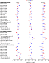
Logarithm of the fold change of free fatty acids (FAs) in each cell line.
Figure 6
Figure 6
Logarithm of the fold change of selected glycerolipids with PUFAs detected in each cell line. PC, phosphatidylcholine; PE, phosphatidylethanolamine; PG, phosphatidylglycerol; LPI, lysophosphatidylinositol; PI, phosphatidylinositol; PS, phosphatidylserine; TG, triacylglyceride.
Figure 7
Figure 7
Logarithm of the fold change of selected glycerolipids with saturated and monounsaturated fatty acids in each cell line. FA, free fatty acids; PC, phosphatidylcholine; PE, phosphatidylethanolamine; TG, triacylglyceride.
Figure 8
Figure 8
Doxorubicin (DOX) treatment increased markers of endoplasmic reticulum stress. (A) Representative images of HepG2, Huh7, and SNU449 tumor cells stained with antibodies against Hoechst stain (blue), DNA damage-inducible transcript 3 (CHOP, red), and binding immunoglobulin protein (BIP, green). (B) Quantification of the levels of CHOP and BIP in the cells with vehicle controls (light red) and treatments with DOX (dark red).
Figure 9
Figure 9
Doxorubicin (DOX) treatment increases the expression of transferrin receptor. (A) Percentage of transferrin receptor-positive staining in the liver of the mouse model untreated (white), induced with hepatocellular carcinoma (HCC, light red), and treated with DOX after induction of HCC (dark red). (B) Representative images of the treatments at large scale (upper row) and detailed scale (lower row). Five replicates were performed; the bar represents the average, and the error bars represent the standard error of the mean.
Scheme 2
Scheme 2
Key molecules in the biosynthetic route of PUFAs and ether glycerolipids in different human subcellular compartments. Adapted from the text in [44,45,46].
Scheme 3
Scheme 3
On the left, schematic interplay between DOX, the lipidome, and ER stress for a cell type, with the output of sensitivity to the drug and cell death mode. On the right, ensemble of qualitative trends of DOX treatment for the three cell lines: (i) the balance between PUFA lipids versus saturated fatty acids (SFAs) and cholesterol lipids (Chol. Lipids), (ii) ER stress, (iii) cell size and the global lipidome, (iv) suggested death mode (Ferr for ferroptosis) and DOX sensitivity.

Similar articles

Cited by

References

    1. Cairns R.A., Harris I.S., Mak T.W. Regulation of Cancer Cell Metabolism. Nat. Rev. Cancer. 2011;11:85–95. doi: 10.1038/nrc2981. - DOI - PubMed
    1. Beloribi-Djefaflia S., Vasseur S., Guillaumond F. Lipid Metabolic Reprogramming in Cancer Cells. Oncogenesis. 2016;5:e189. doi: 10.1038/oncsis.2015.49. - DOI - PMC - PubMed
    1. Kim D.J., Cho E.J., Yu K.-S., Jang I.-J., Yoon J.-H., Park T., Cho J.-Y. Comprehensive Metabolomic Search for Biomarkers to Differentiate Early Stage Hepatocellular Carcinoma from Cirrhosis. Cancers. 2019;11:1497. doi: 10.3390/cancers11101497. - DOI - PMC - PubMed
    1. Smith M.R., Chacko B.K., Johnson M.S., Benavides G.A., Uppal K., Go Y.-M., Jones D.P., Darley-Usmar V.M. A Precision Medicine Approach to Defining the Impact of Doxorubicin on the Bioenergetic-Metabolite Interactome in Human Platelets. Redox Biol. 2020;28:101311. doi: 10.1016/j.redox.2019.101311. - DOI - PMC - PubMed
    1. Sangineto M., Villani R., Cavallone F., Romano A., Loizzi D., Serviddio G. Lipid Metabolism in Development and Progression of Hepatocellular Carcinoma. Cancers. 2020;12:1419. doi: 10.3390/cancers12061419. - DOI - PMC - PubMed

Publication types