

In Vitro Evaluation of Structural Factors Favouring Bacterial Adhesion on Orthodontic Adhesive Resins
- PMID: 34064903
- PMCID: PMC8150295
- DOI: 10.3390/ma14102485
In Vitro Evaluation of Structural Factors Favouring Bacterial Adhesion on Orthodontic Adhesive Resins
Abstract
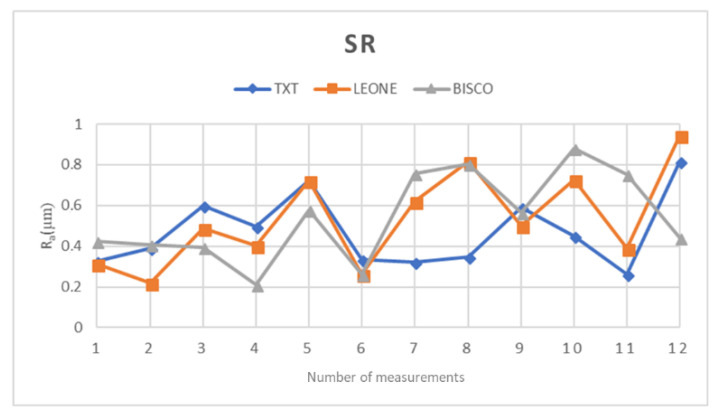
Bacterial adhesion to the surface of orthodontic materials is an important step in the formation and proliferation of plaque bacteria, which is responsible for enamel demineralization and periodontium pathologies. With the intent of investigating if adhesive resins used for bracket bonding are prone to bacteria colonization, the surface roughness of these materials has been analyzed, combining information with a novel methodology to observe the internal structures of orthodontic composites. Scanning electron microscopy, combined with focus ion bean micromachining and stylus profilometry analyses, were performed to evaluate the compositional factors that can influence specific pivotal properties facilitating the adhesion of bacteria to the surface, such as surface roughness and robustness of three orthodontic adhesive composite resins. To confirm these findings, contact angle measurements and bacteria incubation on resin slide have been performed, evaluating similarities and differences in the final achievement. In particular, the morphological features that determine an increase in the resins surface wettability and influence the bacterial adhesion are the subject of speculation. Finally, the focused ion beam technique has been proposed as a valuable tool to combine information coming from surface roughness with specific the internal structures of the polymers.
Keywords: FIB/SEM analysis; bacteria adhesion; orthodontic adhesive resin; surface roughness test.
Conflict of interest statement
The authors declare no conflict of interest.
Figures


Similar articles
-
Influence of surface roughness on streptococcal adhesion forces to composite resins.Dent Mater. 2011 Aug;27(8):770-8. doi: 10.1016/j.dental.2011.03.017. Epub 2011 Apr 27. Dent Mater. 2011. PMID: 21524789
-
Analysis of surface roughness and surface free energy characteristics of various orthodontic materials.Am J Orthod Dentofacial Orthop. 2009 Nov;136(5):668-74. doi: 10.1016/j.ajodo.2007.11.032. Am J Orthod Dentofacial Orthop. 2009. PMID: 19892283
-
Effect of orthodontic debonding and residual adhesive removal on 3D enamel microroughness.PeerJ. 2016 Oct 11;4:e2558. doi: 10.7717/peerj.2558. eCollection 2016. PeerJ. 2016. PMID: 27761343 Free PMC article.
-
Microbial growth on the surfaces of various orthodontic bonding cements.Br J Orthod. 1994 May;21(2):125-32. doi: 10.1179/bjo.21.2.125. Br J Orthod. 1994. PMID: 8043560 Review.
-
Surface properties of resin-based composite materials and biofilm formation: A review of the current literature.Am J Dent. 2015 Dec;28(6):311-20. Am J Dent. 2015. PMID: 26846036 Review.
Cited by
-
Antibiotic Abuse and Antimicrobial Resistance in Hospital Environment: A Retrospective Observational Comparative Study.Medicina (Kaunas). 2022 Sep 11;58(9):1257. doi: 10.3390/medicina58091257. Medicina (Kaunas). 2022. PMID: 36143934 Free PMC article.
-
Physical/Mechanical and Antibacterial Properties of Orthodontic Adhesives Containing Calcium Phosphate and Nisin.J Funct Biomater. 2021 Dec 10;12(4):73. doi: 10.3390/jfb12040073. J Funct Biomater. 2021. PMID: 34940552 Free PMC article.
-
Physical/mechanical and antibacterial properties of orthodontic adhesives containing Sr-bioactive glass nanoparticles, calcium phosphate, and andrographolide.Sci Rep. 2022 Apr 22;12(1):6635. doi: 10.1038/s41598-022-10654-6. Sci Rep. 2022. PMID: 35459791 Free PMC article.
-
Physical and chemical mechanisms involved in adhesion of orthodontic bonding composites: in vitro evaluations.BMC Oral Health. 2021 Jul 16;21(1):350. doi: 10.1186/s12903-021-01715-9. BMC Oral Health. 2021. PMID: 34271907 Free PMC article.
-
Effects of Acidic Environments on Dental Structures after Bracket Debonding.Int J Mol Sci. 2022 Dec 9;23(24):15583. doi: 10.3390/ijms232415583. Int J Mol Sci. 2022. PMID: 36555225 Free PMC article.
References
LinkOut - more resources
Full Text Sources
Molecular Biology Databases
