Review
. 2021 May 11;11(5):1262.
doi: 10.3390/nano11051262.

Peptide-Based Electrospun Fibers: Current Status and Emerging Developments

Affiliations
Review

Peptide-Based Electrospun Fibers: Current Status and Emerging Developments

Raffaella Bucci et al. Nanomaterials (Basel). .

Abstract

Electrospinning is a well-known, straightforward, and versatile technique, widely used for the preparation of fibers by electrifying a polymer solution. However, a high molecular weight is not essential for obtaining uniform electrospun fibers; in fact, the primary criterion to succeed is the presence of sufficient intermolecular interactions, which function similar to chain entanglements. Some small molecules able to self-assemble have been electrospun from solution into fibers and, among them, peptides containing both natural and non-natural amino acids are of particular relevance. Nowadays, the use of peptides for this purpose is at an early stage, but it is gaining more and more interest, and we are now witnessing the transition from basic research towards applications. Considering the novelty in the relevant processing, the aim of this review is to analyze the state of the art from the early 2000s on. Moreover, advantages and drawbacks in using peptides as the main or sole component for generating electrospun nanofibers will be discussed. Characterization techniques that are specifically targeted to the produced peptide fibers are presented.

Keywords: electrospinning; peptide-based electrospun nanofibers; peptides; peptidomimetics; self-assembly.


Conflict of interest statement

The authors declare no conflict of interest.

Figures

Figure 1
Figure 1
Schematic representation of peptide fiber formation via electrospinning.
Figure 2
Figure 2
Phe-Phe electrospun nanofibers of (A) Phe-Phe self-assembled nanotubes of (B). Reproduced from [24] with permission from John Wiley and Sons.
Figure 3
Figure 3
Band-like morphology of Fmoc-Phe-Gly electrospun nanofibers. Reproduced from [41] with permission from The Royal Society of Chemistry.
Figure 4
Figure 4
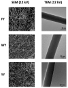
Scanning and transmission electron micrographs SEM and TEM micrographs of Phe-Tyr (FY), Trp-Tyr (WY) and Tyr-Tyr (YY). Reproduced from [25] with permission from The Royal Society of Chemistry.
Figure 5
Figure 5
Chemical structures of compounds 1 and 2.
Figure 6
Figure 6
SEM electron micrographs of electrospun nanofibers of compound 1 (A) and compound 2 (B). Reproduced from [59] with permission from the ACS (https://pubs.acs.org/doi/10.1021/bm401877s (accessed on 10 May 2021)). Further permissions related to the material excerpted should be addressed to the ACS.
Figure 7
Figure 7
Chemical formulae of peptidomimetics 37.
Figure 8
Figure 8
SEM images of electrospun fibers of compound 3. Reproduced from [27] with permission from The Royal Society of Chemistry.
Figure 9
Figure 9
(A) Chemical formula of modified γ-PGA 8. (B) Electrospun materials of compound 8. (C) Strong adhesion of water droplets (“inversion test”). Reproduced from [21] with permission from John Wiley and Sons.
Scheme 1
Scheme 1
Synthetic pathway of PBG and PBGA.
Figure 10
Figure 10
SEM photos of 3D scaffolds of poly(glutamates) fabricated from isotropic fibers (top) and aligned fibers (bottom). Reproduced from [82] with permission from John Wiley and Sons.
Figure 11
Figure 11
SEM images of electrospun PEG/PPGly fibers (A,B) and PPGly fibers after annealing and washing of PEG (C,D). Reproduced from [90] with permission from Wiley-VCH.
Figure 12
Figure 12
Micro- and nanoscale characterization methods. SEM, scanning electron microscopy; EDX, electron-dispersive X-ray spectroscopy; IR, infrared spectroscopy; Raman and fluorescence spectroscopy; XRD, X-ray diffraction; AFM, atomic force microscopy.
Figure 13
Figure 13
Vibrational spectroscopic analysis of Fmoc-Phe-Gly powder (black traces) and the corresponding electrospun fiber (red traces). IR spectrum of (A) powder, (B) fiber, Raman spectrum of (C) fiber and (D) powder. IR was measured in ATR configuration on a diamond prism. Raman scattering was excited at 532 nm. Reproduced from [23] with permission by Wiley-VCH.
Figure 14
Figure 14
AFM analysis of ultrathin electrospun ovalbumin. Courtesy of Dr. A. Eleta Dr. W. Nuansing, CIC nanoGUNE.

Similar articles

Cited by

References

    1. Hosoyama K., Lazurko C., Muñoz M., McTiernan C.D., Alarcon E.I. Peptide-Based Functional Biomaterials for Soft-Tissue Repair. Front. Bioeng. Biotechnol. 2019;7:205. doi: 10.3389/fbioe.2019.00205. - DOI - PMC - PubMed
    1. Chow D., Nunalee M.L., Lim D.W., Simnick A.J., Chilkoti A. Peptide-based biopolymers in biomedicine and biotechnology. Mater. Sci. Eng. R Reports. 2008;62:125–155. doi: 10.1016/j.mser.2008.04.004. - DOI - PMC - PubMed
    1. Reneker D.H., Yarin A.L. Electrospinning jets and polymer nanofibers. Polymer. 2008;49:2387–2425. doi: 10.1016/j.polymer.2008.02.002. - DOI
    1. Singer J.C., Ringk A., Giesa R., Schmidt H.-W. Melt Electrospinning of Small Molecules. Macromol. Mater. Eng. 2015;300:259–276. doi: 10.1002/mame.201400296. - DOI
    1. Raspa A., Pugliese R., Maleki M., Gelain F. Recent therapeutic approaches for spinal cord injury. Biotechnol. Bioeng. 2016;113:253–259. doi: 10.1002/bit.25689. - DOI - PubMed

LinkOut - more resources