

The Impact of Epigenetic Modifications in Myeloid Malignancies
- PMID: 34065087
- PMCID: PMC8125972
- DOI: 10.3390/ijms22095013
The Impact of Epigenetic Modifications in Myeloid Malignancies
Abstract
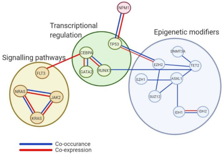
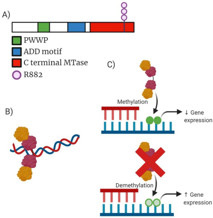
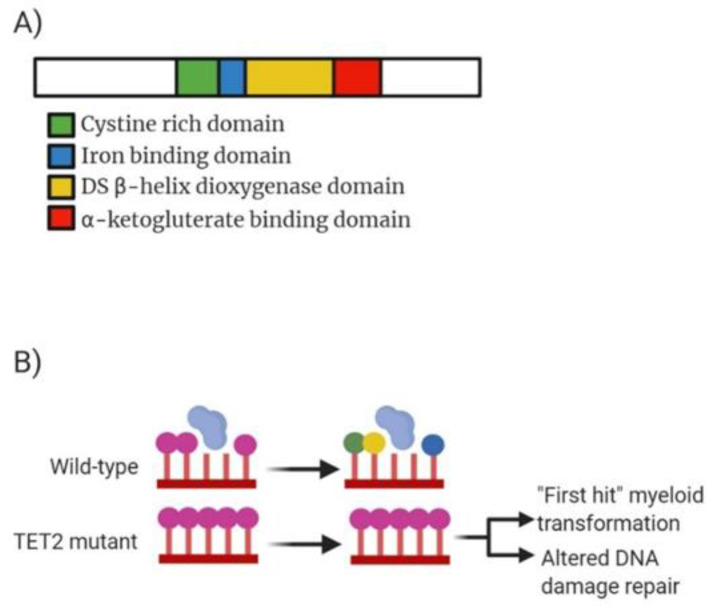
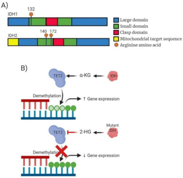
Myeloid malignancy is a broad term encapsulating myeloproliferative neoplasms (MPN), myelodysplastic syndrome (MDS) and acute myeloid leukaemia (AML). Initial studies into genomic profiles of these diseases have shown 2000 somatic mutations prevalent across the spectrum of myeloid blood disorders. Epigenetic mutations are emerging as critical components of disease progression, with mutations in genes controlling chromatin regulation and methylation/acetylation status. Genes such as DNA methyltransferase 3A (DNMT3A), ten eleven translocation methylcytosine dioxygenase 2 (TET2), additional sex combs-like 1 (ASXL1), enhancer of zeste homolog 2 (EZH2) and isocitrate dehydrogenase 1/2 (IDH1/2) show functional impact in disease pathogenesis. In this review we discuss how current knowledge relating to disease progression, mutational profile and therapeutic potential is progressing and increasing understanding of myeloid malignancies.
Keywords: AML; ASXL1; DNMT3A; EZH2; Epigenetics; IDH1; IDH2; MDS; MPN; TET2.
Conflict of interest statement
The authors declare no conflict of interest.
Figures
Similar articles
-
The role of mutations in epigenetic regulators in myeloid malignancies.Nat Rev Cancer. 2012 Sep;12(9):599-612. doi: 10.1038/nrc3343. Epub 2012 Aug 17. Nat Rev Cancer. 2012. PMID: 22898539 Review.
-
[Research on molecular markers for epigenetic changes in myeloid malignancies].Zhonghua Yi Xue Yi Chuan Xue Za Zhi. 2013 Dec;30(6):687-92. doi: 10.3760/cma.j.issn.1003-9406.2013.06.012. Zhonghua Yi Xue Yi Chuan Xue Za Zhi. 2013. PMID: 24327148 Review. Chinese.
-
Mutations in epigenetic regulators in myelodysplastic syndromes.Int J Hematol. 2012 Jan;95(1):8-16. doi: 10.1007/s12185-011-0996-3. Epub 2012 Jan 11. Int J Hematol. 2012. PMID: 22234528 Review.
-
Myeloid malignancies: mutations, models and management.BMC Cancer. 2012 Jul 23;12:304. doi: 10.1186/1471-2407-12-304. BMC Cancer. 2012. PMID: 22823977 Free PMC article. Review.
-
The changing mutational landscape of acute myeloid leukemia and myelodysplastic syndrome.Mol Cancer Res. 2013 Aug;11(8):815-27. doi: 10.1158/1541-7786.MCR-12-0695. Epub 2013 May 3. Mol Cancer Res. 2013. PMID: 23645565 Free PMC article. Review.
Cited by
-
The genesis and evolution of acute myeloid leukemia stem cells in the microenvironment: From biology to therapeutic targeting.Cell Death Discov. 2022 Sep 26;8(1):397. doi: 10.1038/s41420-022-01193-0. Cell Death Discov. 2022. PMID: 36163119 Free PMC article. Review.
-
The roles of phosphorylation of signaling proteins in the prognosis of acute myeloid leukemia.Pathol Oncol Res. 2024 Jul 5;30:1611747. doi: 10.3389/pore.2024.1611747. eCollection 2024. Pathol Oncol Res. 2024. PMID: 39035053 Free PMC article.
-
Methylation of SPRED1: A New Target in Acute Myeloid Leukemia.Front Oncol. 2022 Mar 10;12:854192. doi: 10.3389/fonc.2022.854192. eCollection 2022. Front Oncol. 2022. PMID: 35359401 Free PMC article.
-
DNA methylation-mediated differential expression of DLX4 isoforms has opposing roles in leukemogenesis.Cell Mol Biol Lett. 2022 Jul 26;27(1):59. doi: 10.1186/s11658-022-00358-0. Cell Mol Biol Lett. 2022. PMID: 35883028 Free PMC article.
-
DNA Methyl Transferase 3A (DNMT3A) Mutation Presenting as Isolated Pure Red Cell Aplasia.J Investig Med High Impact Case Rep. 2022 Jan-Dec;10:23247096221097523. doi: 10.1177/23247096221097523. J Investig Med High Impact Case Rep. 2022. PMID: 35593442 Free PMC article.
References
-
- Grimwade D., Hills R.K., Moorman A.V., Walker H., Chatters S., Goldstone A.H., Wheatley K., Harrison C.J., Burnett A.K., on behalf of the National Cancer Research Institute Adult Leukaemia Working Group Refinement of cytogenetic classification in acute myeloid leukemia: Determination of prognostic significance of rare recurring chromosomal abnormalities among 5876 younger adult patients treated in the United Kingdom Medical Research Council trials. Blood. 2010;116:354–365. doi: 10.1182/blood-2009-11-254441. - DOI - PubMed
-
- Fong C.Y., Morison J., Dawson M.A. Epigenetics in the hematologic malignancies [Internet] [(accessed on 7 June 2020)];Haematologica. 2014 997:1772–1783. doi: 10.3324/haematol.2013.092007. Available online: http://www.haematologica.org/content/99/12/1772. - DOI - PMC - PubMed
-
- Abdel-Wahab O., Levine R.L. Mutations in epigenetic modifiers in the pathogenesis and therapy of acute myeloid leukemia. [(accessed on 11 June 2020)];Blood. 2013 121:3563–3572. doi: 10.1182/blood-2013-01-451781. Available online: https://pubmed.ncbi.nlm.nih.gov/23640996/ - DOI - PMC - PubMed
Publication types
MeSH terms
Substances
Grants and funding
LinkOut - more resources
Full Text Sources
Medical
Research Materials
Miscellaneous
