. 2021 May 9;10(5):1141.
doi: 10.3390/cells10051141.

Excessive Iron Induces Oxidative Stress Promoting Cellular Perturbations and Insulin Secretory Dysfunction in MIN6 Beta Cells

Affiliations

Excessive Iron Induces Oxidative Stress Promoting Cellular Perturbations and Insulin Secretory Dysfunction in MIN6 Beta Cells

Voni Blesia et al. Cells. .

Abstract

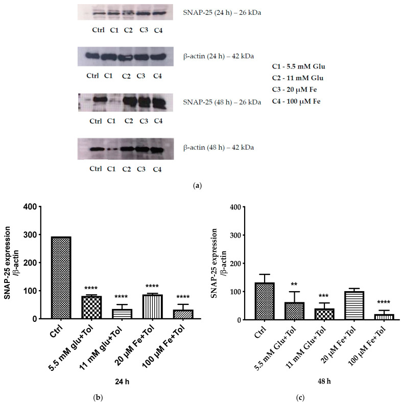
Exposure to high levels of glucose and iron are co-related to reactive oxygen species (ROS) generation and dysregulation of insulin synthesis and secretion, although the precise mechanisms are not well clarified. The focus of this study was to examine the consequences of exposure to high iron levels on MIN6 β-cells. MIN6 pseudoislets were exposed to 20 µM (control) or 100 µM (high) iron at predefined glucose levels (5.5 mM and 11 mM) at various time points (3, 24, 48, and 72 h). Total iron content was estimated by a colourimetric FerroZine™ assay in presence or absence of transferrin-bound iron. Cell viability was assessed by a resazurin dye-based assay, and ROS-mediated cellular oxidative stress was assessed by estimating malondialdehyde levels. β-cell iron absorption was determined by a ferritin immunoassay. Cellular insulin release and content was measured by an insulin immunoassay. Expression of SNAP-25, a key protein in the core SNARE complex that modulates vesicle exocytosis, was measured by immunoblotting. Our results demonstrate that exposure to high iron levels resulted in a 15-fold (48 h) and 4-fold (72 h) increase in cellular iron accumulation. These observations were consistent with data from oxidative stress analysis which demonstrated 2.7-fold higher levels of lipid peroxidation. Furthermore, exposure to supraphysiological (11 mM) levels of glucose and high iron (100 µM) at 72 h exerted the most detrimental effect on the MIN6 β-cell viability. The effect of high iron exposure on total cellular iron content was identical in the presence or absence of transferrin. High iron exposure (100 µM) resulted in a decrease of MIN6 insulin secretion (64% reduction) as well as cellular insulin content (10% reduction). Finally, a significant reduction in MIN6 β-cell SNAP-25 protein expression was evident at 48 h upon exposure to 100 µM iron. Our data suggest that exposure to high iron and glucose concentrations results in cellular oxidative damage and may initiate insulin secretory dysfunction in pancreatic β-cells by modulation of the exocytotic machinery.

Keywords: excess iron; impaired insulin secretion; oxidative stress; type 2 diabetes mellitus; β-cell.


Conflict of interest statement

The authors declare no conflict of interest.

Figures

Figure 1
Figure 1
Effect of transferrin treatments alone or in combination with iron on MIN6 cell intracellular iron content. Data are presented as follows: (a) Dose-response effects of a range of transferrin concentrations (0.005 g/L, 0.05 g/L, 0.5 g/L and 5 g/L) loaded into MIN6 cells for 24 h. The data represent mean ± SEM, n = 3. * p < 0.018. (0.005. g/L vs. 0.5 g/L), * p < 0.033 (0.005 g/L vs. 5 g/L). (b) Effects of 0.05 g/L transferrin loaded into MIN6 cells in combination with 20 μM or 100 μM for 24 h. (c) Effects of 0.5 g/L transferrin loaded into MIN6 cells in combination with 20 μM or 100 μM for 24 h. (d)Effects of 2 g/L transferrin loaded into MIN6 cells in combination with 20 μM or 100 μM for 24 h. (e) Effects of 5 g/L transferrin loaded into MIN6 cells in combination with 20 μM or 100 μM for 24 h. The data (be) represent mean ± SEM, n = 3. Significances are as follows; * p < 0.048 (0.05 g/L),* p < 0.0138 (0.5 g/L), ** p < 0.0036 (2 g/L), ** p < 0.0031 (5 g/L).
Figure 1
Figure 1
Effect of transferrin treatments alone or in combination with iron on MIN6 cell intracellular iron content. Data are presented as follows: (a) Dose-response effects of a range of transferrin concentrations (0.005 g/L, 0.05 g/L, 0.5 g/L and 5 g/L) loaded into MIN6 cells for 24 h. The data represent mean ± SEM, n = 3. * p < 0.018. (0.005. g/L vs. 0.5 g/L), * p < 0.033 (0.005 g/L vs. 5 g/L). (b) Effects of 0.05 g/L transferrin loaded into MIN6 cells in combination with 20 μM or 100 μM for 24 h. (c) Effects of 0.5 g/L transferrin loaded into MIN6 cells in combination with 20 μM or 100 μM for 24 h. (d)Effects of 2 g/L transferrin loaded into MIN6 cells in combination with 20 μM or 100 μM for 24 h. (e) Effects of 5 g/L transferrin loaded into MIN6 cells in combination with 20 μM or 100 μM for 24 h. The data (be) represent mean ± SEM, n = 3. Significances are as follows; * p < 0.048 (0.05 g/L),* p < 0.0138 (0.5 g/L), ** p < 0.0036 (2 g/L), ** p < 0.0031 (5 g/L).
Figure 2
Figure 2
Effect varying of iron and glucose concentrations on intracellular iron content. To quantify the intracellular accumulation of iron, MIN6 cells were exposed to iron (20 μM and 100 μM) and/or glucose (5.5 mM and 11 mM) with the addition of tolbutamide as a secretagogue. Cells were incubated in a time course at 3 and 24 h (a), 48 and 72 h (b). End points measurements were determined using FerroZine™-based colorimetric assay. The data represent mean ± SEM, n = 4. Significance compared to control are as follows ** p < 0.004 (11 mM Glu (C2)—48 h), * p < 0.017 (C3—48 h), *** p < 0.0004 (C4—48 h); * p < 0.014 (5.5 mM Glu (C1)—72 h), *** p < 0.001 (C2—72 h), *** p < 0.0003 (C3—72 h), **** p < 0.0001 (C4—72 h).
Figure 3
Figure 3
The effect of iron (20 μM and 100 μM), glucose (5.5 mM and 11 mM), and combinations of iron and glucose, all with the addition of tolbutamide as a secretagogue on MIN6 cells on ferritin content. This assessment was performed at four different time points [3 and 24 h (a), and 48 and 72 h (b)]. The data represent mean ± SEM, n = 4. Significance compared to control is as follows * p < 0.028 (11 mM Glu (C2)—48 h), * p < 0.023 (20 μM Fe (C3)—48 h), ** p < 0.006 (100 μM Fe (C4)—48 h); * p < 0.025 (C4—72 h).
Figure 4
Figure 4
Determination of lipid peroxidation marker (MDA) performed by TBARS assay (a) following 3 h and 24 h incubation with test conditions; iron (20 μM and 100 μM) and/or glucose (5.5 mM and 11 mM), and combinations of iron and glucose and (b) following 48 h and 72 h incubation with these test conditions (b). High iron and glucose (100 μM and 11 mM respectively) in presence of tolbutamide as a secretagogue increased the concentration of MDA compared to control both at 48 and 72 h time points. The data represent mean ± SEM; n = 4. ** p < 0.0012 (5.5 mM glu—48 h), ** p < 0.001 (11 mM glu—48 h), *** p < 0.0002 (100 μM Fe—48 h), *** p < 0.0003 (5.5 mM glu—72 h), **** p < 0.0001 (11 mM glu and 100 μM Fe—72 h).
Figure 5
Figure 5
The effect of iron and glucose on MIN6 cell viability. Cytotoxicity assessment was performed by PrestoBlue® assay at four incubation times [(3 and 24 h (a), 48 and 72 h (b)]. MIN6 cells were exposed to various concentrations of iron (20 μM and 100 μM) and/or glucose (5.5 mM and 11 mM), and combinations of iron and glucose with the addition of tolbutamide as a secretagogue. The data represent mean ± SEM, n = 4. * p < 0.02 represents significance compared to control.
Figure 6
Figure 6
The effect of iron and glucose on intracellular insulin content. Measurements were performed using insulin ELISA in cultured cells. MIN6 cells were exposed to various concentrations of iron, glucose, and combinations of iron and glucose with the addition of tolbutamide at designated incubation times. The data represent mean ± SEM, n = 3. * p < 0.0193 (20 μM Fe (C3)—12 h), * p < 0.022 (100 μM Fe (C4)—12 h), * p < 0.0148 (11 mM Glu + 100 μM Fe (C8)—12 h); * p < 0.0229 (5.5 mM Glu + 100 uM Fe (C6)—24 h), * p < 0.011 (11 mM Glu + 100 μM Fe (C8)—24 h).
Figure 7
Figure 7
The effect of treatment with varying concentrations of iron, glucose, and combinations of iron and glucose with the addition of tolbutamide on MIN6 cells insulin secretion. (a) The effect of treatment with 5.5 mM and 11 mM of glucose with the addition of tolbutamide on MIN6 cells insulin secretion following 3 h and 24 h incubation. The data represent mean ± SEM, n = 3. (b) The effect of treatment with 20 μM and 100 μM iron with the addition of tolbutamide on MIN6 cells insulin secretion following 3 h and 24 h incubation. The data represent mean ± SEM, n = 3. (c) The effect of treatment with 5.5 mM and 11 mM of glucose and/or 20 μM and 100 μM iron and combinations of iron and glucose with the addition of tolbutamide on MIN6 cells insulin secretion following 3 h and 24 h incubation. The data represent mean ± SEM, n = 3. (d) The effect of treatment incubation with varying concentrations of iron, glucose, and combinations of iron and glucose with the addition of tolbutamide on MIN6 cells insulin secretion acutely (5, 10, 30, 60 min). The data represent mean ± SEM, n = 3. * p < 0.0036 (5.5 mM Glu (C1)—10 min), ** p < 0.037 (11 mM Glu (C2)—10 min), ** p < 0.005 (20 μM Fe (C3)—10 min), * p < 0.026 (5.5 mM Glu + 20 μM Fe (C6)—10 min), * p < 0.01 (11 mM Glu + 100 μM Fe (C8)—10 min); * p < 0.05 (C1—30 min), * p < 0.033 (C2—30 min), * p < 0.016 (C3—30 min), * p < 0.026 (C6—30 min), * p < 0.01 (C8—30 min); * p < 0.013 (C3—60 min). (e) The effect of treatment with varying concentrations of iron, glucose, and combinations of iron and glucose with the addition of tolbutamide on MIN6 cells insulin secretion following 24 h (5, 10, 30, 60 min). The data represent mean ± SEM, n = 3. * p < 0.049 [11 mM Glu (C2)—24 h (5 min)], * p < 0.022 [20 μM Fe (C3)—24 h (5 min)], ** p < 0.004 [100 μM Fe (C4)—24 h (5 min)], * p < 0.013 [5.5 mM Glu + 100 μM Fe—24 h (C6)(5 min)], ** p < 0.003 [11 mM Glu + 100 μM Fe—24 h (C8)(5 min)]; * p < 0.034 [C2—24 h(10 min)], * p < 0.022 [C3—24 h(10 min)], * p < 0.02 (C4—24 h(10 min)], ** p < 0.005 [C6—24 h(10 min)], * p < 0.027 [C8—24 h(10 min)]; * p < 0.015 [C2—24 h(30 min)], ** p < 0.004 [C3—24 h(30 min)], * p < 0.016 [C4—24 h(30 min)], ** p < 0.005 [C6—24 h(30 min)], * p < 0.018 [C8—24 h(30 min)]; * p < 0.044 [C2—24 h(60 min)], * p < 0.034 (C3, C4, C6—24 h(60 min)].
Figure 7
Figure 7
The effect of treatment with varying concentrations of iron, glucose, and combinations of iron and glucose with the addition of tolbutamide on MIN6 cells insulin secretion. (a) The effect of treatment with 5.5 mM and 11 mM of glucose with the addition of tolbutamide on MIN6 cells insulin secretion following 3 h and 24 h incubation. The data represent mean ± SEM, n = 3. (b) The effect of treatment with 20 μM and 100 μM iron with the addition of tolbutamide on MIN6 cells insulin secretion following 3 h and 24 h incubation. The data represent mean ± SEM, n = 3. (c) The effect of treatment with 5.5 mM and 11 mM of glucose and/or 20 μM and 100 μM iron and combinations of iron and glucose with the addition of tolbutamide on MIN6 cells insulin secretion following 3 h and 24 h incubation. The data represent mean ± SEM, n = 3. (d) The effect of treatment incubation with varying concentrations of iron, glucose, and combinations of iron and glucose with the addition of tolbutamide on MIN6 cells insulin secretion acutely (5, 10, 30, 60 min). The data represent mean ± SEM, n = 3. * p < 0.0036 (5.5 mM Glu (C1)—10 min), ** p < 0.037 (11 mM Glu (C2)—10 min), ** p < 0.005 (20 μM Fe (C3)—10 min), * p < 0.026 (5.5 mM Glu + 20 μM Fe (C6)—10 min), * p < 0.01 (11 mM Glu + 100 μM Fe (C8)—10 min); * p < 0.05 (C1—30 min), * p < 0.033 (C2—30 min), * p < 0.016 (C3—30 min), * p < 0.026 (C6—30 min), * p < 0.01 (C8—30 min); * p < 0.013 (C3—60 min). (e) The effect of treatment with varying concentrations of iron, glucose, and combinations of iron and glucose with the addition of tolbutamide on MIN6 cells insulin secretion following 24 h (5, 10, 30, 60 min). The data represent mean ± SEM, n = 3. * p < 0.049 [11 mM Glu (C2)—24 h (5 min)], * p < 0.022 [20 μM Fe (C3)—24 h (5 min)], ** p < 0.004 [100 μM Fe (C4)—24 h (5 min)], * p < 0.013 [5.5 mM Glu + 100 μM Fe—24 h (C6)(5 min)], ** p < 0.003 [11 mM Glu + 100 μM Fe—24 h (C8)(5 min)]; * p < 0.034 [C2—24 h(10 min)], * p < 0.022 [C3—24 h(10 min)], * p < 0.02 (C4—24 h(10 min)], ** p < 0.005 [C6—24 h(10 min)], * p < 0.027 [C8—24 h(10 min)]; * p < 0.015 [C2—24 h(30 min)], ** p < 0.004 [C3—24 h(30 min)], * p < 0.016 [C4—24 h(30 min)], ** p < 0.005 [C6—24 h(30 min)], * p < 0.018 [C8—24 h(30 min)]; * p < 0.044 [C2—24 h(60 min)], * p < 0.034 (C3, C4, C6—24 h(60 min)].
Figure 8
Figure 8
SNAP-25 protein expressions in pancreatic β-cell (MIN6) within two distinct timelines, 24 and 48 h, in the presence of iron (20 μM and 100 μM) and glucose (5.5 mM and 11 mM) with the addition of tolbutamide as a secretagogue. Protein expression was identified using Western blotting. (a) Distinctive bands of SNAP-25 (24 and 48 h) and β-actin (24 and 48 h). Gel loading was as follows (from top left): lane 1—ctrl, lane 2—normal glucose (5.5 mM) (C1), lane 3—high glucose (11 mM) (C2), lane 4—normal iron (20 μM) (C3), lane 5—high iron (100 μM) (C4); (from middle right): lane 1—ctrl, lane 2—C1, lane 3—C2, lane 4—C3, lane 5—C4. (b) Expression of SNAP-25 at 24 h normalised against β-actin. (c) Expression of SNAP-25 at 48 h normalised against β-actin. The data represent mean ± SEM; n = 4. **** p < 0.0001 (24 h), ** p < 0.0061 (48 h), *** p < 0.0005, **** p < 0.0001 (48 h).

Similar articles

Cited by

References

    1. Guariguata L., Whiting D.R., Hambleton I., Beagley J., Linnenkamp U., Shaw J.E. Global Estimates of Diabetes Prevalence for 2013 and Projections for 2035. Diabetes Res. Clin. Pract. 2014;103:137–149. doi: 10.1016/j.diabres.2013.11.002. - DOI - PubMed
    1. Wild S., Roglic G., Green A., Sicree R., King H. Global Prevalence of Diabetes: Estimates for the Year 2000 and Projections for 2030. Diabetes Care. 2004;27:1047–1053. doi: 10.2337/diacare.27.5.1047. - DOI - PubMed
    1. Tripathy D., Chavez A.O. Defects in Insulin Secretion and Action in the Pathogenesis of Type 2 Diabetes Mellitus. Curr. Diab. Rep. 2010;10:184–191. doi: 10.1007/s11892-010-0115-5. - DOI - PubMed
    1. Jewell J.L., Oh E., Thurmond D.C. Exocytosis Mechanisms Underlying Insulin Release and Glucose Uptake: Conserved Roles for Munc18c and Syntaxin 4. Am. J. Physiol. Regul. Integr. Comp. Physiol. 2010;298:R517–R531. doi: 10.1152/ajpregu.00597.2009. - DOI - PMC - PubMed
    1. Gerber P.A., Rutter G.A. The Role of Oxidative Stress and Hypoxia in Pancreatic Beta-Cell Dysfunction in Diabetes Mellitus. Antioxid. Redox Signal. 2017;26:501–518. doi: 10.1089/ars.2016.6755. - DOI - PMC - PubMed

Publication types