Review
. 2021 May 9;9(5):526.
doi: 10.3390/biomedicines9050526.

Alternative Splicing of Human Telomerase Reverse Transcriptase (hTERT) and Its Implications in Physiological and Pathological Processes

Affiliations
Review

Alternative Splicing of Human Telomerase Reverse Transcriptase (hTERT) and Its Implications in Physiological and Pathological Processes

Anna A Plyasova et al. Biomedicines. .

Abstract

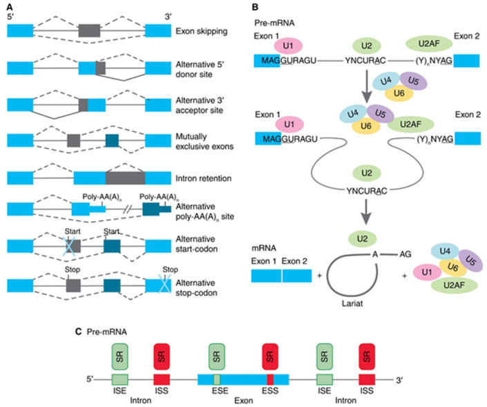
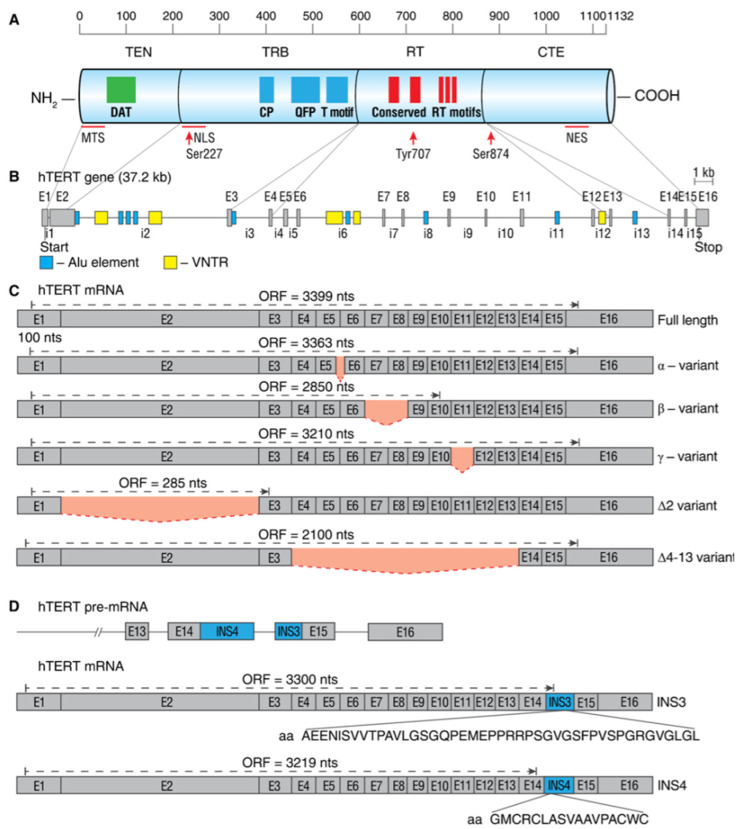
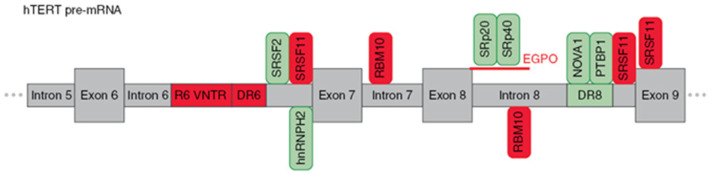
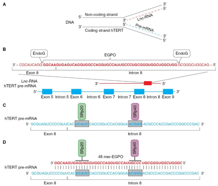
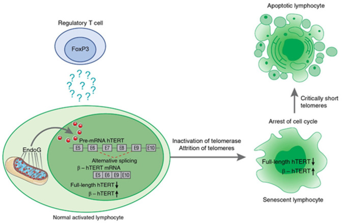
Alternative splicing (AS) of human telomerase catalytic subunit (hTERT, human telomerase reverse transcriptase) pre-mRNA strongly regulates telomerase activity. Several proteins can regulate AS in a cell type-specific manner and determine the functions of cells. In addition to being involved in telomerase activity regulation, AS provides cells with different splice variants that may have alternative biological activities. The modulation of telomerase activity through the induction of hTERT AS is involved in the development of different cancer types and embryos, and the differentiation of stem cells. Regulatory T cells may suppress the proliferation of target human and murine T and B lymphocytes and NK cells in a contact-independent manner involving activation of TERT AS. This review focuses on the mechanism of regulation of hTERT pre-mRNA AS and the involvement of splice variants in physiological and pathological processes.

Keywords: alternative splicing; apoptosis; endonuclease G; human telomerase reverse transcriptase (hTERT); lymphocytes; splice variants; telomerase; telomeres.


Conflict of interest statement

Authors declare no conflict of interests.

Figures

Figure 1
Figure 1
Schematic presentation of biology of alternative splicing (AS). (A) Traditional classification of basic types of alternative RNA splicing events. Exons are represented as blue and grey blocks, introns as lines in between. (B) Functioning of spliceosome. Pre-mRNA containing two exons separated by an intron assembles into splicing complexes together with spliceosome subunits. Individual subunits are indicated by U1, U2, U2AF, U4, U5, and U6. U1 forms a base-pairing interaction with 5′-splice site, whereas U2 base-pairs with branch-point and U2AF binds to polypyrimidine site of 3′ splice site. Then, a complex containing U4, U5, and U6 associates with the forming of spliceosome. The intron is removed in a form of lariat and two exons are ligated. (C) Interactions of trans-elements and serine/arginine-rich (SR) proteins with cis-elements, and regulatory sequences on pre-mRNA. Elements that inhibit exon inclusion are shown in red, while those enhancing inclusion are shown in green. ESE, exonic splicing enhancer; ESI, exonic splicing silencer; ISE, intronic splicing enhancer; ISS, intronic splicing silencer.
Figure 2
Figure 2
Schematic map of hTERT protein, gene, and commonly studied mRNA splice variants. (A) Linear structure of 1132-amino acid hTERT protein and known domains and motifs are shown. The following active elements are responsible for intracellular relocalization of hTERT: MTS, mitochondrial targeting sequences; NLS, nuclear localization signal; Ser227, Serine 227 for phosphorylation by Akt; Tyr770, Tyrosine 770 for phosphorylation by Src1; Ser824, Serine 824 for phosphorylation by Akt; NES, nuclear export signal for binding with CRM1. (B) Structure of hTERT gene exons (E1–E16) and introns (i1–i15). Positions of Alu elements and variable number tandem repeats (VNTRs) are shown as dark blue and yellow boxes, respectively. Lines link exons and the domains they encode. (C) Common alternatively spliced variants with deletions are shown below the wild-type, the full-length mRNA. Predicted open reading frame (ORF) for each mRNA is indicated. (D) Common alternatively spliced variants that include insertions INS3 and INS4 and the amino acids that are encoded by them.
Figure 3
Figure 3
Schematic presentation of cis-elements and trans-factors involved in the regulation of β- alternative splicing of hTERT pre-mRNA. Entire region from intron 5 to exon 9 of hTERT pre-mRNA is shown. Inclusion enhancers of exons 7 and 8 are shown in green, while repressors are shown in red. EndoG-produced oligonucleotides that block binding of two SR proteins are shown in red.
Figure 4
Figure 4
Schematic presentation of mechanism of hTERT pre-mRNA splicing induced by EndoG. (A) Hypothetical locations for synthesis of hTERT pre-mRNA and lnc-RNA. Pre-mRNA hTERT (blue dotted line) is synthesized from coding strand of hTERT gene, while lnc-RNA (red dotted line) is synthesized from the noncoding DNA strand. (B) Hypothetical schematic locations for lncRNAs and EndoG-produced oligonucleotides (EGPOs) during regulation of hTERT pre-mRNA AS by EndoG. EndoG (white box) cleaves EGPO (red bold font) from lnc-RNA (red font), which is complementary to hTERT pre-mRNA. (C) Binding sites (blue bold font in gray boxes) for the SRp20 (green box) and SRp40 (purple box) splicing regulatory proteins are located in intron 8. (D) Interaction between EGPO and hTERT pre-mRNA prevents binding of SRp20 and SRp40 to hTERT pre-mRNA, which results in induction of AS and expression of truncated β- splice variant.
Figure 5
Figure 5
Schematic presentation of Treg-induced mechanism of hTERT pre-mRNA alternative splicing during suppression of target lymphocyte proliferation. Treg cells induce EndoG expression and translocation from mitochondria to nucleus in activated lymphocytes by a contact-independent mechanism. The factor that triggers this process is not yet determined. From its location in nucleus of target lymphocytes, EndoG induces β- splicing, resulting in inhibition of telomerase. Prolonged cocultivation with Tregs leads to telomere attrition, cell cycle arrest, conversion of the target cells to replicative senescence, and apoptotic death.

Similar articles

Cited by

References

    1. Trybek T., Kowalik A., Góźdź S., Kowalska A. Telomeres and telomerase in oncogenesis (review) Oncol. Lett. 2020;20:1015–1027. doi: 10.3892/ol.2020.11659. - DOI - PMC - PubMed
    1. Ozturk S. Telomerase activity and telomere length in male germ cells. Biol. Reprod. 2015;92:53. doi: 10.1095/biolreprod.114.124008. - DOI - PubMed
    1. Hiyama E., Hiyama K. Telomere and telomerase in stem cells. Br. J. Cancer. 2007;96:1020–1024. doi: 10.1038/sj.bjc.6603671. - DOI - PMC - PubMed
    1. Akbar A.N., Vukmanovic-Stejic M. Telomerase in T lymphocytes: Use it and lose it? J. Immunol. 2007;178:6689–6694. doi: 10.4049/jimmunol.178.11.6689. - DOI - PubMed
    1. Jakob S., Haendeler J. Molecular mechanisms involved in endothelial cell aging: Role of telomerase reverse transcriptase. Z. Gerontol. Geriatr. 2007;40:334–338. doi: 10.1007/s00391-007-0482-y. - DOI - PubMed