

RABL6A Regulates Schwann Cell Senescence in an RB1-Dependent Manner
- PMID: 34065204
- PMCID: PMC8161079
- DOI: 10.3390/ijms22105367
RABL6A Regulates Schwann Cell Senescence in an RB1-Dependent Manner
Abstract
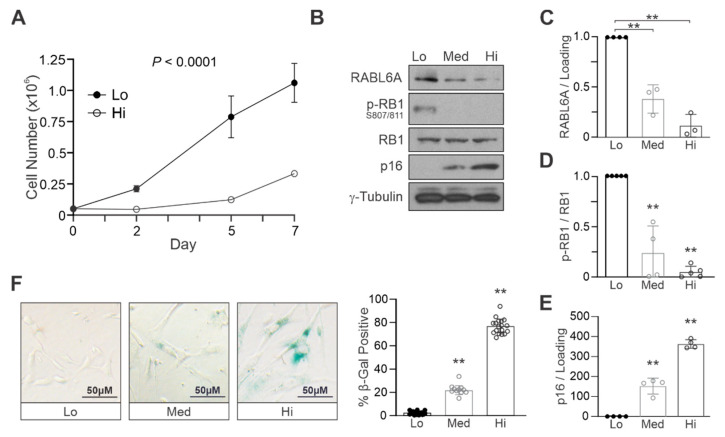
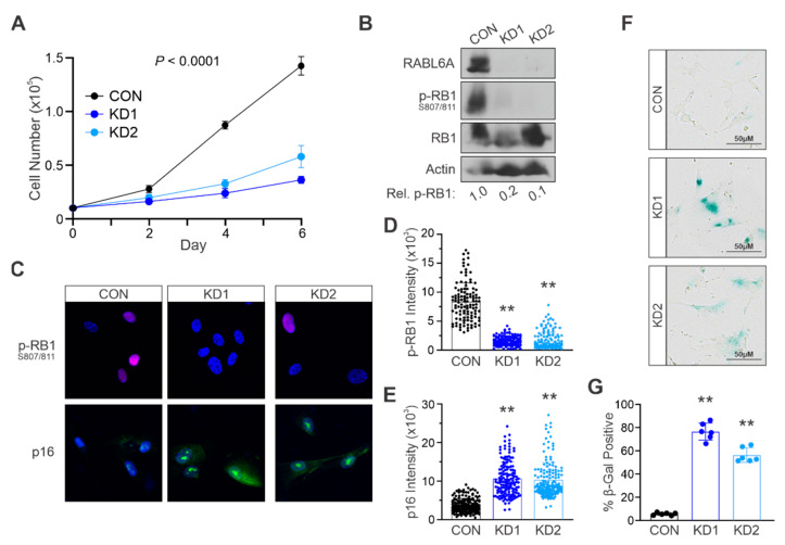
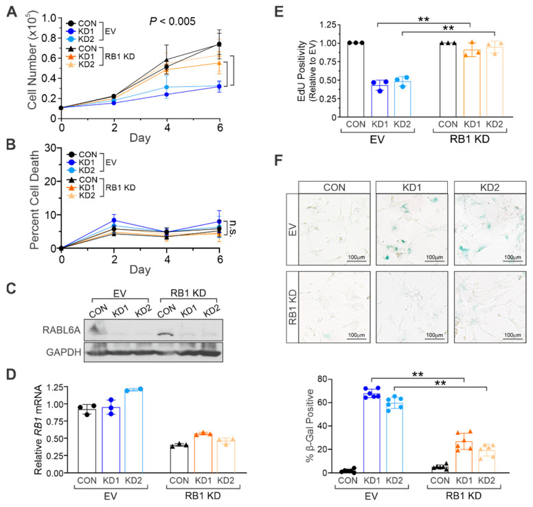
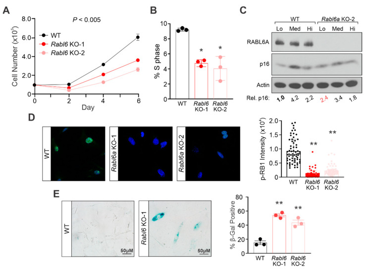
Schwann cells are normally quiescent, myelinating glia cells of the peripheral nervous system. Their aberrant proliferation and transformation underlie the development of benign tumors (neurofibromas) as well as deadly malignant peripheral nerve sheath tumors (MPNSTs). We discovered a new driver of MPNSTs, an oncogenic GTPase named RABL6A, that functions in part by inhibiting the RB1 tumor suppressor. RB1 is a key mediator of cellular senescence, a permanent withdrawal from the cell cycle that protects against cell immortalization and transformation. Based on the RABL6A-RB1 link in MPNSTs, we explored the hypothesis that RABL6A promotes Schwann cell proliferation and abrogates their senescence by inhibiting RB1. Using sequentially passaged normal human Schwann cells (NHSCs), we found that the induction of replicative senescence was associated with reduced expression of endogenous RABL6A. Silencing RABL6A in low passage NHSCs caused premature stress-induced senescence, which was largely rescued by co-depletion of RB1. Consistent with those findings, Rabl6-deficient MEFs displayed impaired proliferation and accelerated senescence compared to wildtype MEFs. These results demonstrate that RABL6A is required for maintenance of proper Schwann cell proliferation and imply that aberrantly high RABL6A expression may facilitate malignant transformation.
Keywords: MPNST; RABL6A; Schwann cell; retinoblastoma protein (RB1); senescence.
Conflict of interest statement
The authors declare no conflict of interest.
Figures
Similar articles
-
RABL6A Is an Essential Driver of MPNSTs that Negatively Regulates the RB1 Pathway and Sensitizes Tumor Cells to CDK4/6 Inhibitors.Clin Cancer Res. 2020 Jun 15;26(12):2997-3011. doi: 10.1158/1078-0432.CCR-19-2706. Epub 2020 Feb 21. Clin Cancer Res. 2020. PMID: 32086342 Free PMC article.
-
RABL6A promotes G1-S phase progression and pancreatic neuroendocrine tumor cell proliferation in an Rb1-dependent manner.Cancer Res. 2014 Nov 15;74(22):6661-70. doi: 10.1158/0008-5472.CAN-13-3742. Epub 2014 Oct 1. Cancer Res. 2014. PMID: 25273089 Free PMC article.
-
Combination therapies for MPNSTs targeting RABL6A-RB1 signaling.Oncotarget. 2021 Jan 5;12(1):10-14. doi: 10.18632/oncotarget.27862. eCollection 2021 Jan 5. Oncotarget. 2021. PMID: 33456709 Free PMC article.
-
The Challenge of Cancer Genomics in Rare Nervous System Neoplasms: Malignant Peripheral Nerve Sheath Tumors as a Paradigm for Cross-Species Comparative Oncogenomics.Am J Pathol. 2016 Mar;186(3):464-77. doi: 10.1016/j.ajpath.2015.10.023. Epub 2015 Dec 28. Am J Pathol. 2016. PMID: 26740486 Free PMC article. Review.
-
RB1: a prototype tumor suppressor and an enigma.Genes Dev. 2016 Jul 1;30(13):1492-502. doi: 10.1101/gad.282145.116. Genes Dev. 2016. PMID: 27401552 Free PMC article. Review.
Cited by
-
Novel Therapeutics and the Path Toward Effective Immunotherapy in Malignant Peripheral Nerve Sheath Tumors.Cancers (Basel). 2025 Jul 21;17(14):2410. doi: 10.3390/cancers17142410. Cancers (Basel). 2025. PMID: 40723291 Free PMC article. Review.
-
m5C-dependent cross-regulation between nuclear reader ALYREF and writer NSUN2 promotes urothelial bladder cancer malignancy through facilitating RABL6/TK1 mRNAs splicing and stabilization.Cell Death Dis. 2023 Feb 18;14(2):139. doi: 10.1038/s41419-023-05661-y. Cell Death Dis. 2023. PMID: 36806253 Free PMC article.
-
From benign neurofibromas to malignant peripheral nerve sheath tumors (MPNST): a gaming among multiple factors.Cell Oncol (Dordr). 2025 Aug;48(4):841-857. doi: 10.1007/s13402-025-01054-9. Epub 2025 Apr 2. Cell Oncol (Dordr). 2025. PMID: 40172801 Free PMC article. Review.
-
FOXM1, MEK, and CDK4/6: New Targets for Malignant Peripheral Nerve Sheath Tumor Therapy.Int J Mol Sci. 2023 Sep 2;24(17):13596. doi: 10.3390/ijms241713596. Int J Mol Sci. 2023. PMID: 37686402 Free PMC article. Review.
-
The Role of CDK Pathway Dysregulation and Its Therapeutic Potential in Soft Tissue Sarcoma.Cancers (Basel). 2022 Jul 12;14(14):3380. doi: 10.3390/cancers14143380. Cancers (Basel). 2022. PMID: 35884441 Free PMC article. Review.
References
-
- Kidd G.J., Ohno N., Trapp B.D. Biology of Schwann cells. Handb. Clin. Neurol. 2013;115:55–79. - PubMed
MeSH terms
Substances
Grants and funding
- Young Investigator Award/Children's Tumor Foundation
- P30 ES005605/ES/NIEHS NIH HHS/United States
- Synodos for Neurofibromatosis-1/Children's Tumor Foundation
- P30 CA086862/CA/NCI NIH HHS/United States
- Sarcoma Multidisciplinary Oncology Group Pilot Award/Holden Comprehensive Cancer Center, University of Iowa
LinkOut - more resources
Full Text Sources
Molecular Biology Databases
Miscellaneous
