. 2021 May 20;22(10):5405.
doi: 10.3390/ijms22105405.

Curcumin at Low Doses Potentiates and at High Doses Inhibits ABT-737-Induced Platelet Apoptosis

Affiliations

Curcumin at Low Doses Potentiates and at High Doses Inhibits ABT-737-Induced Platelet Apoptosis

Natalia Rukoyatkina et al. Int J Mol Sci. .

Abstract

Curcumin is a natural bioactive component derived from the turmeric plant Curcuma longa, which exhibits a range of beneficial activities on human cells. Previously, an inhibitory effect of curcumin on platelets was demonstrated. However, it is unknown whether this inhibitory effect is due to platelet apoptosis or procoagulant platelet formation. In this study, curcumin did not activate caspase 3-dependent apoptosis of human platelets, but rather induced the formation of procoagulant platelets. Interestingly, curcumin at low concentration (5 µM) potentiated, and at high concentration (50 µM) inhibited ABT-737-induced platelet apoptosis, which was accompanied by inhibition of ABT-737-mediated thrombin generation. Platelet viability was not affected by curcumin at low concentration and was reduced by 17% at high concentration. Furthermore, curcumin-induced autophagy in human platelets via increased translocation of LC3I to LC3II, which was associated with activation of adenosine monophosphate (AMP) kinase and inhibition of protein kinase B activity. Because curcumin inhibits P-glycoprotein (P-gp) in cancer cells and contributes to overcoming multidrug resistance, we showed that curcumin similarly inhibited platelet P-gp activity. Our results revealed that the platelet inhibitory effect of curcumin is mediated by complex processes, including procoagulant platelet formation. Thus, curcumin may protect against or enhance caspase-dependent apoptosis in platelets under certain conditions.

Keywords: apoptosis; autophagy; platelets; procoagulant activity; thrombin.


Conflict of interest statement

The authors declare no competing interests.

Figures
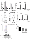
Figure 1
Figure 1
Curcumin inhibits thrombin-induced platelet αIIbβ3 integrin activation and does not stimulate caspase 3-dependent apoptosis. (A) Flow cytometric analysis of αIIbβ3 integrin activation (PAC-1-FITC binding), (B) PS surface exposure (annexin-V-PE binding), (C,D) microparticle formation, (E,F) mitochondrial membrane potential changes (TMRE fluorescence), and (G) Western blot of caspase 3 activation. Washed platelets (WP 1 × 10⁸ /mL in A-F and 3 × 10⁸/mL in G) were incubated with the indicated concentrations of curcumin for 10 and 60 min. (A) Thrombin (0.01 U/mL) was added for 2 min, followed by PAC-1-FITC antibody (1:10 dilution) for 10 min, and the reaction was stopped by dilution (10 volumes) with PBS. (B) WP were incubated with the indicated concentrations/time of curcumin, then annexin-V-PE (dilution 1:10) was added for an additional 10 min, and the reaction was stopped by dilution (10 volumes) with the annexin-V-binding solution. (C) Representative (from four independent experiments) dot plot of microparticle formation (upper panel), and annexin-V-PE positive platelets and microparticles (lower panel). Annexin-V-PE was analyzed as shown in B. (D) Quantification of platelet microparticle formation. Microparticles were quantified as CD42a positive events in the gate B. (E,F) WP were incubated with curcumin (50 µM, 10 and 60 min), TMRE dye (dilution 1:10) was added for an additional 10 min, and samples were diluted (10 volumes) with PBS. (G) WP were incubated with the indicated concentrations/time of curcumin and processed for Western blotting with caspase 3 antibody (1:1000). ABT-737 was used as positive control and actin blot served as a loading control. All data are presented as means ± SD. Data in A are presented as % of MFI (thrombin sample represents 100%, one-way ANOVA, Levene’s test p > 0.05 followed by Tukey’s HSD test, * p = 0.0001 compared to controls, n = 5). In B, as % of annexin-V positive platelets (one-way ANOVA, Levene’s test p > 0.05 followed by Tukey’s HSD test, * p = 0.0001 compared to controls, n = 5). In D, as fold increase of microparticles (control taken as 1), one-way ANOVA, Levene’s test p < 0.05 followed by Tamhane’s T2 test, * p = 0.0001 compared to controls, n = 5). In F, as % of TMRE fluorescence intensity change (control represents 100%, one-way ANOVA, Levene’s test p > 0.05 followed by Tukey’s HSD test, * p < 0.05, n.s.—not significant, n = 6).
Figure 2
Figure 2
Curcumin increases doxorubicin accumulation in platelets. Washed platelets (1 × 10⁸ /mL) were incubated with the indicated concentrations of curcumin alone or in combination with doxorubicin (20 µM) for 60 min in the absence (control) and analyzed by flow cytometry for doxorubicin fluorescence. P-gp inhibitor cyclosporin A was used as a positive control. (A) Representative dot plots, (B) quantification of six independent experiments (curcumin autofluorescence was subtracted from 50 µM curcumin samples). Data are presented as means ± SD (One-way ANOVA, Levene’s test p < 0.05 followed by Tamhane’s T2 test, * p = 0.01 compared to control, n = 4).
Figure 3
Figure 3
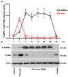
Curcumin stimulates AMPK and inhibits PKB activity in platelets. Washed platelets (3 × 10⁸ cells/mL) were incubated with curcumin (50 µM) for the indicated time and analyzed by Western blotting for total actin, AMPK, and PKB phosphorylation. Thrombin (0.01 U/mL, 1 min) was used as a positive control for PKB and AMPK phosphorylation. (A) Quantitative analysis of PKB and AMPK phosphorylation. Immunoblots were scanned and quantified by the Image J program. The intensity of the P-AMPK and P-PKB signal was normalized to the actin signal. For each sample, this ratio is relatively expressed to the ratio for control, which is presented as 1. Data are presented as means ± SD. One-way ANOVA, Levene’s test p > 0.05 followed by Tukey’s HSD test for P-AMPK and Levene’s test p < 0.05 followed by Tamhane’s T2 test for P-PKB. For P-AMPK * as appeared in the figure p = 0.0001; 0.0001; 0.0001; 0.0001; 0.0001, n = 4. For P-PKB * p = 0.049; 0.012; 0.02; 0.001 (n = 5), compared to controls taken as 1 in both cases. (B) Representative Western blots of PKA and AMPK phosphorylation.
Figure 4
Figure 4
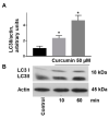
Curcumin induces the conversion of LC3I to LC3II in platelets. Washed platelets (3 × 10⁸ /mL) were incubated with curcumin (50 µM) for the indicated time and analyzed by Western blotting for LC3 translocation (LC3I/LC3II). (A) Immunoblots were scanned and the intensity of bands was quantified by the ImageJ program. The intensity of the LC3/II signal was normalized to the actin signal. For each sample, this is relatively expressed to the ratio for the control, which is presented as 1. Data are presented as means ± SD. Data are presented as means ± SD. One-way ANOVA, Levene’s test p < 0.05 followed by Tamhane’s T2 test, * as appeared in the figure p = 0.002; p = 0.019, n = 5, compared to controls taken as 1. (B) Representative Western blot of LC3 translocation.
Figure 5
Figure 5
Curcumin had no effect on ABT-737-induced phosphatidylserine surface exposure. Washed platelets (1 × 10⁸ /mL) were incubated with indicated concentrations of ABT-737 in the absence or presence of curcumin (5, 50 µM) and PS-positive platelets (annexin-V-PE binding) were analyzed by flow cytometry, Data are presented as means ± SD, n = 6. Paired t-test p = 0.52, p = 0.13; and one-way ANOVA, Levene’s test p > 0.05 followed by Tukey’s HSD test, p = 0.678, p = 0.871, n.s.—not significant.
Figure 6
Figure 6
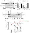
Curcumin at low concentrations stimulated and at high concentration inhibited platelet apoptosis induced by low concentrations of ABT-737. (A,C) Washed platelets (3 × 10⁸ cells/mL) were incubated with the indicated concentrations of curcumin and ABT-737 for 60 min and analyzed by Western blotting for caspase 3 activation (procaspase 3, cleaved caspase 3). Actin blot served as a loading control. (B,D) Immunoblots were scanned and the intensity of bands was quantified by the ImageJ program. The intensity of the procaspase 3 signal was normalized to the actin signal. For each sample, this ratio is relatively expressed to the ratio for the control, which is presented as 1. Data are presented as means ± SD. For 6B: One-way ANOVA, Levene’s test p > 0.05 followed by Tukey’s HSD test * as appeared in the figure, p = 0.017; 0.001; 0.002; 0.005 compared to control, presented as 1. For 6D: One-way ANOVA, Levene’s test p > 0.05 followed by Tukey’s HSD test, compared to the corresponding controls, * as appeared in the figure p = 0.005; 0.002; # as appeared in the figure p = 0.001; 0.001; Paired t-test (“ABT-737 + Curcumin” vs. “ABT-737”) $ as appeared in the figure, p = 0.004; 0.002; n = 4.
Figure 7
Figure 7
Curcumin at high concentration enhances thrombin-triggered and abolishes ABT-737-mediated thrombin generation on human platelets. Washed platelets (9 × 10⁸ cells/mL) were preincubated with curcumin (5 µM/C5, 50 µM/C50) and/or ABT-737 (0.5 µM) for 60 min, adjusted to 1.5 × 10⁸/mL with autologous platelet-free plasma. Platelet-dependent thrombin generation capacity triggered by α-thrombin (0.1 U/mL) was monitored by calibrated automated thrombography. (A) Representative thrombogram curves. (B) Quantitative thrombin generation data expressed as thrombin peak in % compared to thrombin control. (C) Quantitative data of annexin-V-PE binding to washed human platelets in platelet-free plasma induced by 0.1 U/mL α-thrombin in the absence or presence of curcumin and/or ABT-737, expressed as a percentage of annexin-V-PE positive platelets. Data are presented as means ± SD of 3 separate experiments from 3 different donors (One-way ANOVA, Levene’s test p > 0.05 followed by Tukey’s HSD test), n.s. not significant.

Similar articles

Cited by

References

    1. Baell J.B. Feeling nature′s PAINS: Natural products, natural product drugs, and Pan Assay Interference Compounds (PAINS) J. Nat. Prod. 2016;79:616–628. doi: 10.1021/acs.jnatprod.5b00947. - DOI - PubMed
    1. Reddy A.C., Lokesh B.R. Effect of dietary turmeric (Curcuma longa) on iron-induced lipid peroxidation in the rat liver. Food Chem. Toxicol. 1994;32:279–283. doi: 10.1016/0278-6915(94)90201-1. - DOI - PubMed
    1. Yang M., Akbar U., Mohan C. Curcumin in autoimmune and rheumatic diseases. Nutrients. 2019;11:1004. doi: 10.3390/nu11051004. - DOI - PMC - PubMed
    1. He Y., Yue Y., Zheng X., Zhang K., Chen S., Du Z. Curcumin, inflammation, and chronic diseases: How are they linked? Molecules. 2015;20:9183–9213. doi: 10.3390/molecules20059183. - DOI - PMC - PubMed
    1. Farhood B., Mortezaee K., Goradel N.H., Khanlarkhani N., Salehi E., Nashtaei M.S., Najafi M., Sahebkar A. Curcumin as an anti-inflammatory agent: Implications to radiotherapy and chemotherapy. J. Cell Physiol. 2019;234:5728–5740. doi: 10.1002/jcp.27442. - DOI - PubMed

MeSH terms