Review
. 2021 May 20;22(10):5407.
doi: 10.3390/ijms22105407.

An Analytical Review of the Structural Features of Pentatricopeptide Repeats: Strategic Amino Acids, Repeat Arrangements and Superhelical Architecture

Affiliations
Review

An Analytical Review of the Structural Features of Pentatricopeptide Repeats: Strategic Amino Acids, Repeat Arrangements and Superhelical Architecture

Sailen Barik. Int J Mol Sci. .

Abstract

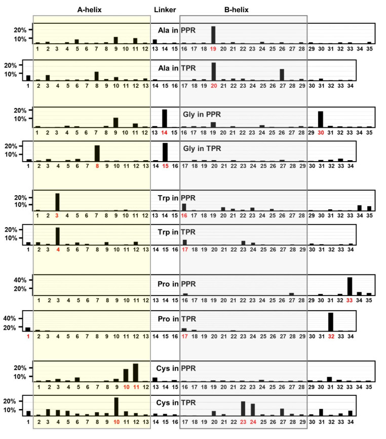
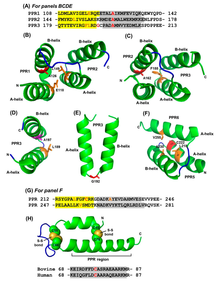
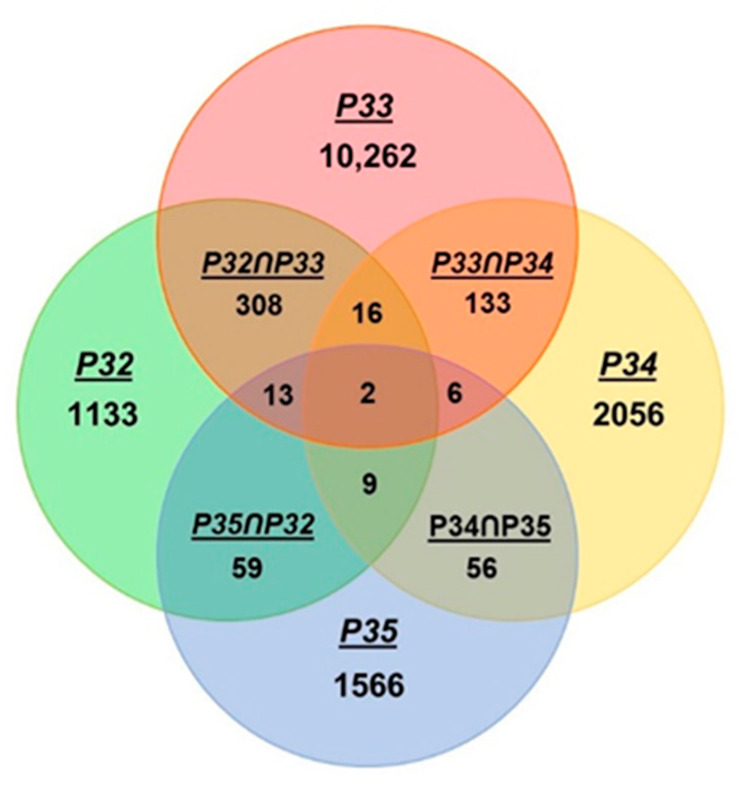
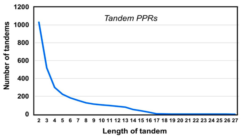
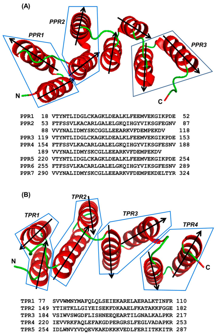
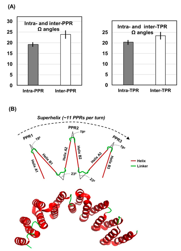
Tricopeptide repeats are common in natural proteins, and are exemplified by 34- and 35-residue repeats, known respectively as tetratricopeptide repeats (TPRs) and pentatricopeptide repeats (PPRs). In both classes, each repeat unit forms an antiparallel bihelical structure, so that multiple such units in a polypeptide are arranged in a parallel fashion. The primary structures of the motifs are nonidentical, but amino acids of similar properties occur in strategic positions. The focus of the present work was on PPR, but TPR, its better-studied cousin, is often included for comparison. The analyses revealed that critical amino acids, namely Gly, Pro, Ala and Trp, were placed at distinct locations in the higher order structure of PPR domains. While most TPRs occur in repeats of three, the PPRs exhibited a much greater diversity in repeat numbers, from 1 to 30 or more, separated by spacers of various sequences and lengths. Studies of PPR strings in proteins showed that the majority of PPR units are single, and that the longer tandems (i.e., without space in between) occurred in decreasing order. The multi-PPR domains also formed superhelical vortices, likely governed by interhelical angles rather than the spacers. These findings should be useful in designing and understanding the PPR domains.

Keywords: PPR; helix; protein structure; protein-RNA interaction; solvation; tricopeptide repeats.


Conflict of interest statement

The authors declare no conflict of interest.

Figures

Figure 1
Figure 1
Positional distribution of individual amino acids over penta- and tetra-tricopeptide repeats. Naturally occurring tetra- and penta-tricopeptide repeat sequences (a total of 22,999 PPRs and 1137 TPRs) retrieved as described previously [10] and in the Materials and Methods. For each amino acid, the total number in each position of the repeat is counted using Excel, and plotted as the percentage of the total number of that amino acid in all positions (35 in PPR and 34 in TPR). For example, the percentage of Ala at any PPR position is = (Na/Nt) × 100, where Na is the number of Ala at a given position a in all PPRs, and Nt is total number of Ala in all positions in all PPRs. Note the Y-axis scale for Pro is higher because the bulk of it (~50%) is present at a single position (as Pro33/Pro32), which in fact serves as a signature for these repeats [2,3,4,10]. Numbers in red on the X-axis indicate the major site(s) of concentration of a residue. A-helix and B-helix are boxed in transparent yellow and grey, respectively, with the linker sequence in the middle.
Figure 2
Figure 2
Interaction between the side chains of selective amino acid residues in pentatricopeptide repeats (PPRs). The 3D structures displayed by PyMol in panels (BF,H) correspond to the amino acid sequences in panels (A,G,H), as labeled (details in Results). A-helix and B-helix sequences are shaded yellow and grey, respectively. The sequence in panel (A) is a set of three-PPR repeats in the Arabidopsis thaliana ‘thylakoid assembly 8-like protein’ found in the chloroplast (GenBank Q9STF9; PDB 4LEU). In each repeat, the invariant A20 residue and a glycine are in red color, and the major residues that the interact with are in orange, as shown in the panels (BE). Panel (G) shows two tandem PPRs in proteinaceous Ribonuclease P1, also from a six-PPR domain in Arabidopsis thaliana chloroplast (Q66GI4), corresponding to the structure in panel (F) (PDB 4G24). Panel (H) shows the part of a unique CHCH/PPR domain conserved in bovine and human, which is involved in two pairs of intramolecular Cys-Cys disulfide bonds as shown (details in Results, Section 2.1.5).
Figure 3
Figure 3
Combinatorial locations of Pro residues in PPR. The occurrence of Pro at the distal end of PPR was counted by Excel’s ‘countif’ function as described in Results (Section 2.1.4). Colocalizations of Pro at multiple sites were also counted similarly and the numbers presented in the overlapping areas of the Euler diagram (which may appear similar to the more commonly known ‘Venn diagram’).
Figure 4
Figure 4
Arrangements of PPR. The graph presents the relative abundance of PPR clusters of different lengths. For example, 3 in the X-axis means a cluster of three tandem PPR units connected without any spacer amino acid between them, i.e., PPR-PPR-PPR. As seen in the graph, the PPR-PPR-PPR triplet was found 519 times in our collection of 2130 non-redundant PPR-containing proteins (details in Section 2.2).
Figure 5
Figure 5
Superhelical structure of a multi-PPR domain. The superhelical structure of a PPR domain is shown along with that of a TPR domain to illustrate the similarities. (A) PPR domain of the plant ‘multiple organellar RNA editing factor’ (MORF, also known as ‘RNA editing factor interacting protein’ or RIP) from PDB 5IWW, Chain D [41]. The first three PPRs (out of a total of seven) are shown as representative, and demarcated by polygons. The two unboxed helices belong to a non-PPR sequence between the second and third PPR. (B) TPR domain (for comparison) of RRP5, an essential factor for ribosome maturation in yeast; PDB WWM, Chain B [24]. Only four TPRs of the multi-TPR C-terminal region are shown and each is demarcated by a polygon. In both panels A and B, the helices are dark red in color and the connecting linkers are green. The two termini of the peptide (amino, N and carboxy, C) are labeled, and the directionality of each helix is also indicated by arrowheads, which shows their antiparallel arrangement.
Figure 6
Figure 6
Helical angles in PPR domains. (A) Interhelical angles (Ω) determined from PDB crystal structures; the averages of 208 intra-PPR, 234 inter-PPR, and 216 each of intra- and inter-TPR helical angles are shown. The exact values and the standard errors are in parenthesis: intra-PPR (19.22 ± 1.37), inter-PPR (23.9 ± 3.03); intra-TPR (20.36 ± 1.63), inter-TPR (23.41 ± 3.27). (B) Schematic of location of the inter-and inter-repeat helical angles are located in the context of a superhelix, which was found to contain approximately 11 PPRs per complete turn. For a realistic perspective, an actual superhelix of a multi-TPR domain (PDB 5C9S, a portion of which was shown in Figure 5B) is shown below. Note that the superhelices of PPR and TPR are virtually identical.
Figure 7
Figure 7
Single-helix (unihelical) TPR units. (A) Only the first four TPR sequences of the seven-TPR T. brucei PRX5 protein are aligned to mark the similar amino acids in TPR1, TPR2 and one TPR3 (shaded in grey, if present in two or more TPRs) while the general TPR signature residues are shown on top. The A-helix and the B-helix are colored red; note that the two helices of TPR3 are discernible in the structure of the full-length protein that contain all seven TPRs (PDB structure 3CV0), but form one continuous long helix in the truncated protein (PDB structure 1HXI), indicated by the continuous red font color. (B) The first three TPR units of the 3CV0 (red color) and 1HXI (cyan color), corresponding to the sequences in Panel A, are superimposed using the merge function of PyMol. Note the bihelical structure of TPR3 in 3CV0 (red), and unihelical structure of the same TPR in 1HXI (cyan); TPR1 and TPR2 have retained the same bihelical structures in both constructs, as seen by complete superimposition. (C) Comparison of the helical regions, observed by X-ray diffraction of crystals of the full-length protein phosphatase 5 (PP5) and those of the 3-TPR domain only, devoid of the downstream sequence (PDB 1WAO and 1A17, respectively). Only the TPR3 (shaded grey) and the relevant portion of the downstream sequence are shown, as TPR1 and TPR2 structures were very similar in the two crystals. All helices—TPR and non-TPR—are colored red. Note that the shorter fragment of PP5 has the canonical bihelical architecture, whereby the LGK loop region separates the A- and B-helices (not marked); the TPR is followed by a long 35-aa helix (KDA…VDS). In contrast, the full-length protein (1WAO) shows a unihelical structure of the TPR, followed by a shorter helix, only 19-aa long (DAK..AFE).

Similar articles

Cited by

References

    1. Kajava A.V. Tandem repeats in proteins: From sequence to structure. J. Struct. Biol. 2012;179:279–288. doi: 10.1016/j.jsb.2011.08.009. - DOI - PubMed
    1. Sawyer N., Chen J., Regan L. All repeats are not equal: A module-based approach to guide repeat protein design. J. Mol. Biol. 2013;425:1826–1838. doi: 10.1016/j.jmb.2013.02.013. - DOI - PMC - PubMed
    1. Manna S. An overview of pentatricopeptide repeat proteins and their applications. Biochimie. 2015;113:93–99. doi: 10.1016/j.biochi.2015.04.004. - DOI - PubMed
    1. Small I.D., Peeters N. The PPR motif—A TPR-related motif prevalent in plant organellar proteins. Trends Biochem. Sci. 2000;25:46–47. doi: 10.1016/S0968-0004(99)01520-0. - DOI - PubMed
    1. Kobayashi K., Kawabata M., Hisano K., Kazama T., Matsuoka K., Sugita M., Nakamura T. Identification and characterization of the RNA binding surface of the pentatricopeptide repeat protein. Nucleic Acids Res. 2012;40:2712–2723. doi: 10.1093/nar/gkr1084. - DOI - PMC - PubMed

LinkOut - more resources