. 2021 May 16;13(5):921.
doi: 10.3390/v13050921.

Amino Acid Substitutions in NS5 Contribute Differentially to Tembusu Virus Attenuation in Ducklings and Cell Cultures

Affiliations

Amino Acid Substitutions in NS5 Contribute Differentially to Tembusu Virus Attenuation in Ducklings and Cell Cultures

Xue Sun et al. Viruses. .

Abstract

Tembusu virus (TMUV), a highly infectious pathogenic flavivirus, causes severe egg-drop and encephalitis in domestic waterfowl, while the determinants responsible for viral pathogenicity are largely unknown. In our previous studies, virulent strain JXSP2-4 had been completely attenuated by successive passages in BHK-21 cells and the avirulent strain was designated as JXSP-310. Based on the backbone of JXSP2-4, a series of chimeric viruses were generated according to the amino acid substitutions in NS5 and their infectivities were also analyzed in cell cultures and ducklings. The results showed that the viral titers of RNA-dependent RNA polymerase (RdRp) domain-swapped cheimeric mutant (JXSP-310RdRp) in cells and ducklings were both markedly decreased compared with JXSP2-4, indicating that mutations in the RdRp domain affected viral replication. There are R543K and V711A two amino acid substitutions in the RdRp domain. Further site-directed mutagenesis showed that single-point R543K mutant (JXSP-R543K) exhibited similar replication efficacy compared with JXSP2-4 in cells, but the viral loads in JXSP-R543K-infected ducklings were significantly lower than that of JXSP2-4 and higher than JXSP-310RdRp. Surprisingly, the single-point V711A mutation we introduced rapidly reverted. In addition, qRT-PCR and Western blot confirmed that the mutations in the RdRp domain significantly affected the replication of the virus. Taken together, these results show that R543K substitution in the RdRp domain impairs the in vivo growth of TMUV, but sustaining its attenuated infectivity requires the concurrent presence of the V711A mutation.

Keywords: RNA-dependent RNA polymerase; Tembusu virus; attenuation; chimeric viruses; viral replication.


Conflict of interest statement

The authors declare no conflict of interest.

Figures

Figure 1
Figure 1
Characteristics of chimeric viruses in vitro. (A) Strategy used to construct chimeric and mutant viruses. The JXSP-310MTase chimera contains two mutations (V153 and S226), JXSP-310Linker contains two mutations (R273, G277) and JXSP-310RdRp also contains two mutations (R543, V711). The filled circles represent the parental JXSP2-4 (orange) and JXSP-310 (blue) viruses, respectively. (B) Immunofluorescence assay. BHK-21 cells were fixed after 48 h infection with the chimeric viruses and stained with an antibody against the E protein. Scale bar 100 μm. (C) Plaque phenotypes of JXSP2-4 (n = 50) and JXSP-310RdRp (n = 50). The mean ± SD values are shown and the data were tested for statistical significance using the Student’s t test. ***, p < 0.001. (D) Multi-step growth curves of the chimeric viruses. BHK-21 cells and DEF cells were separately infected with JXSP2-4, JXSP-310MTase, JXSP-310Linker or JXSP-310RdRp, each at an MOI of 0.1. The culture supernatants were collected every 12 h and viral titers were quantified using plaque assay. All experiments were performed in triplicate and the mean ± the SD values are shown.
Figure 2
Figure 2
The viral loads of the chimeric viruses at 2 day post-infection. Seven-day-old ducklings were subcutaneously inoculated with JXSP2-4, JXSP-310MTase, JXSP-310Linker and JXSP-310RdRp (1 × 10⁵ PFU/bird) separately and euthanized 2 days later. Viral loads in the blood (A), spleen (B), heart (C), kidneys (D), lungs (E), liver (F), thymus (G) and bursa of Fabricius (H) were determined using plaque assay in BHK-21 cells. Data were tested for statistical significance by two-way multiple ANOVA comparisons. Each chimeric virus was compared with JXSP2-4 (**, p < 0.05; ***, p < 0.001; ****, p < 0.0001). The dashed line represents the detection limit of the assay.
Figure 3
Figure 3
The viral loads of JXSP2-4 and JXSP-310RdRp at various time points. Seven-day-old ducklings were inoculated with JXSP2-4 and JXSP-310RdRp (1×10⁵ PFU/bird) and then euthanized at day 1, 2, 4 and 6 post-infection. Viral loads in the blood (A), spleen (B), heart (C), kidneys (D), lungs (E), liver (F), thymus (G) and bursa of Fabricius (H) were determined using plaque assay in BHK-21 cells. The data were tested for statistical significance by two-way multiple ANOVA comparisons (*, p < 0.05; **, p < 0.01; ***, p < 0.001; ****, p < 0.0001). The dashed line represents the detection limit of the assay.
Figure 4
Figure 4
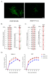
Characterization of mutant viruses in vitro. (A) Immunofluorescence assay. BHK-21 cells were fixed at 48 h after infection with either of the rescue viruses and stained with antibody against the E protein. Scale bar 100 μm. (B,C) Whole genome sequence analysis of 10 serially passaged mutant viruses. The JXSP-R543K mutant virus was stable during serial passage, but the 711-site mutant reverted to Val (GTA). (D) Multi-step growth curves of the mutant viruses. BHK-21 cells and DEF cells were infected with JXSP2-4, JXSP-310RdRp and JXSP-R543K (MOI of 0.1). The culture supernatants were collected every 12 h and the viral titers were quantified using plaque assay. All experiments were performed in triplicate and the mean ± SD values are shown.
Figure 5
Figure 5
The viral loads of JXSP-R543K, JXSP-310RdRp and JXSP2-4 at various time points. Seven-day-old ducklings were inoculated with JXSP2-4, JXSP-310RdRp and JXSP-R543K (1 × 10⁵ PFU/bird) separately and euthanized at days 1, 2 and 4 post-infection. Viral titers in the blood (A), spleen (B), heart (C), kidneys (D), lungs (E), liver (F), thymus (G) and bursa of Fabricius (H) were determined using plaque assay in BHK-21 cells. Asterisks indicate significant differences between JXSP2-4 and JXSP-R543K, (*, p < 0.05; **, p < 0.01; ***, p < 0.001; ****, p < 0.0001). Number signs indicate significant differences between JXSP-310RdRp and JXSP-R543K (#, p < 0.05; ##, p < 0.01; ###, p < 0.001; ####, p < 0.0001). The data were tested for statistical significance by two-way multiple ANOVA comparisons. The dashed line represents the detection limit of the assay.
Figure 6
Figure 6
Immune-related gene expression among JXSP2-4-, JXSP-310RdRp-and JXSP-R543K-infected ducklings. The mRNA levels of IFN-α, IFN-β, IFN-γ, IL-1β and IL-6 in the spleens were analyzed by qRT-PCR and normalized to GADPH. Asterisks indicate significant differences between JXSP2-4 and JXSP-R543K, (*, p < 0.05; **, p < 0.01; ***, p < 0.001; ****, p < 0.0001). Number signs indicate significant differences between JXSP-310RdRp and JXSP-R543K (##, p < 0.01; ####, p < 0.0001). The data were tested for statistical significance by two-way multiple ANOVA comparisons.
Figure 7
Figure 7
RNA synthesis and protein production of the viruses. (A) Quantitative analysis of RNA production in the parental virus, chimeric viruses and the single-site mutant virus by qRT-PCR in DEF cells. (B,C) Protein lysates from DEF cells collected at 24 hpi and 36 hpi were analyzed by western blotting using antibodies against the E protein and the internal β-actin control. The numbers represent the ratios in comparison with JXSP2-4, which were determined by densitometry.
Figure 8
Figure 8
Details of the side-chain interactions between residues R543 and V711 in TMUV RdRp. (A) The structure of NS5 RdRp of TMUV shows the fingers, palm and thumb domains colored in blue, grey and orange, respectively. The TMUV NS5 protein model was created online using Swiss-Model with reference to the crystal structure of the JEV NS5 protein (PDB identifier [ID] 4k6m). (B) R543 is located in motif A and possibly forms three solid hydrogen bonds with W540, S690 and K691 spatially. V711 is close to motif E and forms one solid hydrogen bond with W708. (C) An amino acid mutation at site 543 changes arginine to lysine, which breaks the hydrogen bond between sites 543 and 690. A valine to alanine change at site 711 slightly changes the distance between motif A and residue W708. This figure was generated in PyMol.

Similar articles

Cited by

References

    1. Liu P., Lu H., Li S., Moureau G., Deng Y.Q., Wang Y., Zhang L., Jiang T., de Lamballerie X., Qin C.F., et al. Genomic and antigenic characterization of the newly emerging Chinese duck egg-drop syndrome flavivirus: Genomic comparison with Tembusu and Sitiawan viruses. J. Gen. Virol. 2012;93:2158–2170. doi: 10.1099/vir.0.043554-0. - DOI - PubMed
    1. Benzarti E., Linden A., Desmecht D., Garigliany M. Mosquito-borne epornitic flaviviruses: An update and review. J. Gen. Virol. 2019;100:119–132. doi: 10.1099/jgv.0.001203. - DOI - PubMed
    1. Ninvilai P., Tunterak W., Oraveerakul K., Amonsin A., Thontiravong A. Genetic characterization of duck Tembusu virus in Thailand, 2015–2017: Identification of a novel cluster. Transbound. Emerg. Dis. 2019;66:1982–1992. doi: 10.1111/tbed.13230. - DOI - PubMed
    1. Mackenzie J.S., Williams D.T. The zoonotic flaviviruses of southern, south-eastern and eastern Asia, and Australasia: The potential for emergent viruses. Zoonoses Public Health. 2009;56:338–356. doi: 10.1111/j.1863-2378.2008.01208.x. - DOI - PubMed
    1. Yan P., Zhao Y., Zhang X., Xu D., Dai X., Teng Q., Yan L., Zhou J., Ji X., Zhang S., et al. An infectious disease of ducks caused by a newly emerged Tembusu virus strain in mainland China. Virology. 2011;417:1–8. doi: 10.1016/j.virol.2011.06.003. - DOI - PubMed

Publication types

Supplementary concepts