

Recent Advancements in Enzyme-Based Lateral Flow Immunoassays
- PMID: 34065971
- PMCID: PMC8150770
- DOI: 10.3390/s21103358
Recent Advancements in Enzyme-Based Lateral Flow Immunoassays
Abstract
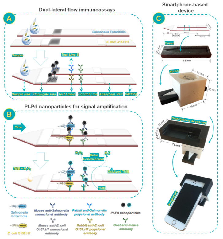
Paper-based lateral-flow immunoassays (LFIAs) have achieved considerable commercial success and their impact in diagnostics is continuously growing. LFIA results are often obtained by visualizing by the naked eye color changes in given areas, providing a qualitative information about the presence/absence of the target analyte in the sample. However, this platform has the potential to provide ultrasensitive quantitative analysis for several applications. Indeed, LFIA is based on well-established immunological techniques, which have known in the last year great advances due to the combination of highly sensitive tracers, innovative signal amplification strategies and last-generation instrumental detectors. All these available progresses can be applied also to the LFIA platform by adapting them to a portable and miniaturized format. This possibility opens countless strategies for definitively turning the LFIA technique into an ultrasensitive quantitative method. Among the different proposals for achieving this goal, the use of enzyme-based immunoassay is very well known and widespread for routine analysis and it can represent a valid approach for improving LFIA performances. Several examples have been recently reported in literature exploiting enzymes properties and features for obtaining significative advances in this field. In this review, we aim to provide a critical overview of the recent progresses in highly sensitive LFIA detection technologies, involving the exploitation of enzyme-based amplification strategies. The features and applications of the technologies, along with future developments and challenges, are also discussed.
Keywords: chemiluminescence; colorimetric; enzyme; lateral flow immunoassay; metal nanoparticles; nanozyme; point-of-care.
Conflict of interest statement
The authors declare no conflict of interest.
Figures

Similar articles
-
Nanocellulose aerogel inserts for quantitative lateral flow immunoassays.Biosens Bioelectron. 2021 Nov 15;192:113491. doi: 10.1016/j.bios.2021.113491. Epub 2021 Jul 9. Biosens Bioelectron. 2021. PMID: 34271399
-
Automatic ultrasensitive lateral flow immunoassay based on a color-enhanced signal amplification strategy.Biosens Bioelectron. 2024 Jul 15;256:116262. doi: 10.1016/j.bios.2024.116262. Epub 2024 Mar 30. Biosens Bioelectron. 2024. PMID: 38621340
-
Field detection capability of immunochemical assays during criminal investigations involving the use of TNT.Forensic Sci Int. 2015 Jan;246:25-30. doi: 10.1016/j.forsciint.2014.10.037. Epub 2014 Oct 31. Forensic Sci Int. 2015. PMID: 25460104
-
A nanoparticle-assisted signal-enhancement technique for lateral flow immunoassays.J Mater Chem B. 2024 Jul 17;12(28):6735-6756. doi: 10.1039/d4tb00865k. J Mater Chem B. 2024. PMID: 38920348 Review.
-
Advances in enhancement-type signal tracers and analysis strategies driven Lateral flow immunoassay for guaranteeing the agri-food safety.Biosens Bioelectron. 2025 Jan 15;268:116920. doi: 10.1016/j.bios.2024.116920. Epub 2024 Nov 7. Biosens Bioelectron. 2025. PMID: 39531800 Review.
Cited by
-
Lateral flow assay: a promising rapid point-of-care testing tool for infections and non-communicable diseases.Asian Biomed (Res Rev News). 2023 Dec 28;17(6):250-266. doi: 10.2478/abm-2023-0068. eCollection 2023 Dec. Asian Biomed (Res Rev News). 2023. PMID: 38161347 Free PMC article. Review.
-
Enhancing a SARS-CoV-2 nucleocapsid antigen test sensitivity with cost efficient strategy through a cotton intermembrane insertion.Sci Rep. 2023 Mar 22;13(1):4690. doi: 10.1038/s41598-023-31641-5. Sci Rep. 2023. PMID: 36949174 Free PMC article.
-
An Origami Paper-Based Biosensor for Allergen Detection by Chemiluminescence Immunoassay on Magnetic Microbeads.Biosensors (Basel). 2022 Oct 4;12(10):825. doi: 10.3390/bios12100825. Biosensors (Basel). 2022. PMID: 36290961 Free PMC article.
-
Highly Sensitive Lateral Flow Immunodetection of the Insecticide Imidacloprid in Fruits and Berries Reached by Indirect Antibody-Label Coupling.Foods. 2024 Dec 25;14(1):25. doi: 10.3390/foods14010025. Foods. 2024. PMID: 39796315 Free PMC article.
-
Optimizing the Composition of the Substrate Enhances the Performance of Peroxidase-like Nanozymes in Colorimetric Assays: A Case Study of Prussian Blue and 3,3'-Diaminobenzidine.Molecules. 2023 Nov 16;28(22):7622. doi: 10.3390/molecules28227622. Molecules. 2023. PMID: 38005344 Free PMC article.
References
-
- Mak W.C., Beni V., Turner A.P. Lateral-flow technology: From visual to instrumental. TrAC Trends Anal. Chem. 2016;79:297–305. doi: 10.1016/j.trac.2015.10.017. - DOI
-
- Mahmoudi T., de la Guardia M., Baradaran B. Lateral flow assays towards point-of-care cancer detection: A review of cur-rent progress and future trends. TrAC-Trend Anal. Chem. 2020;125:115842. doi: 10.1016/j.trac.2020.115842. - DOI
Publication types
MeSH terms
Grants and funding
LinkOut - more resources
Full Text Sources
Other Literature Sources
